Bioscience COPD/IPF, Research & Early Development, Respiratory & Immunology, BioPharmaceuticals R&D, AstraZeneca, Cambridge, UK.
Translational Sciences & Experimental Medicine, Research & Early Development, Respiratory & Immunology, BioPharmaceuticals R&D, AstraZeneca, Gothenburg, Sweden.
Respir Res. 2023 Feb 14;24(1):51. doi: 10.1186/s12931-023-02333-5.
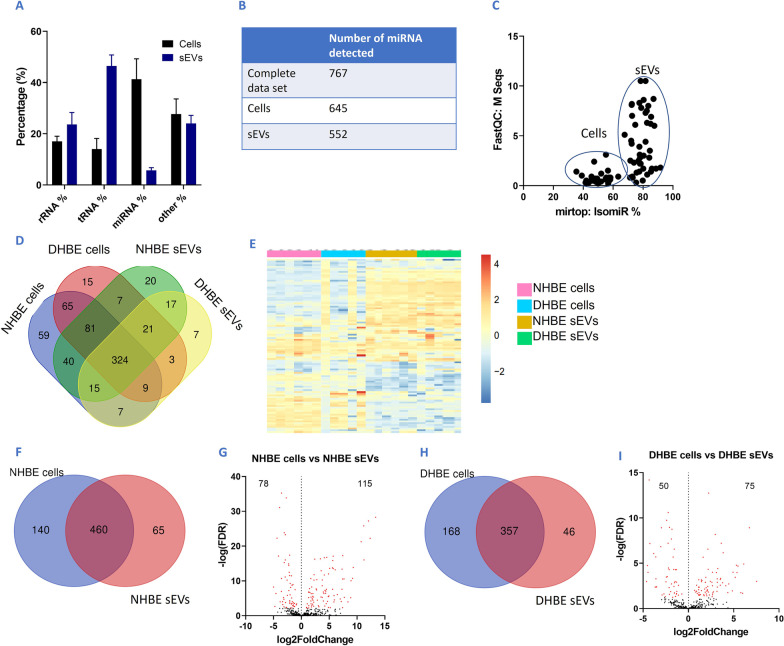
Idiopathic pulmonary fibrosis (IPF) is a chronic lung disease that affects 3 million people worldwide. Senescence and small extracellular vesicles (sEVs) have been implicated in the pathogenesis of IPF, although how sEVs promote disease remains unclear. Here, we profile sEVs from bronchial epithelial cells and determine small RNA (smRNA) content.
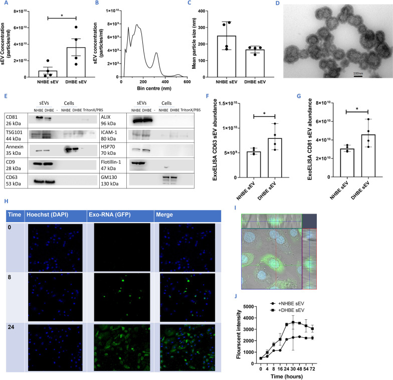
Conditioned media was collected and sEVs were isolated from normal human bronchial epithelial cells (NHBEs) and IPF-diseased human bronchial epithelial cells (DHBEs).
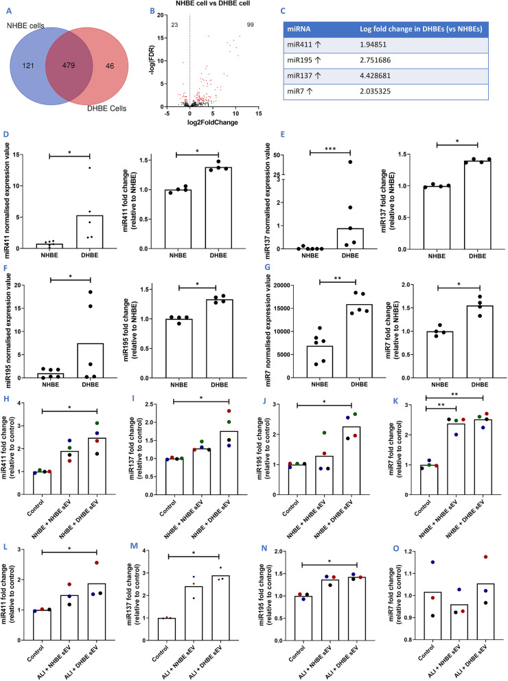
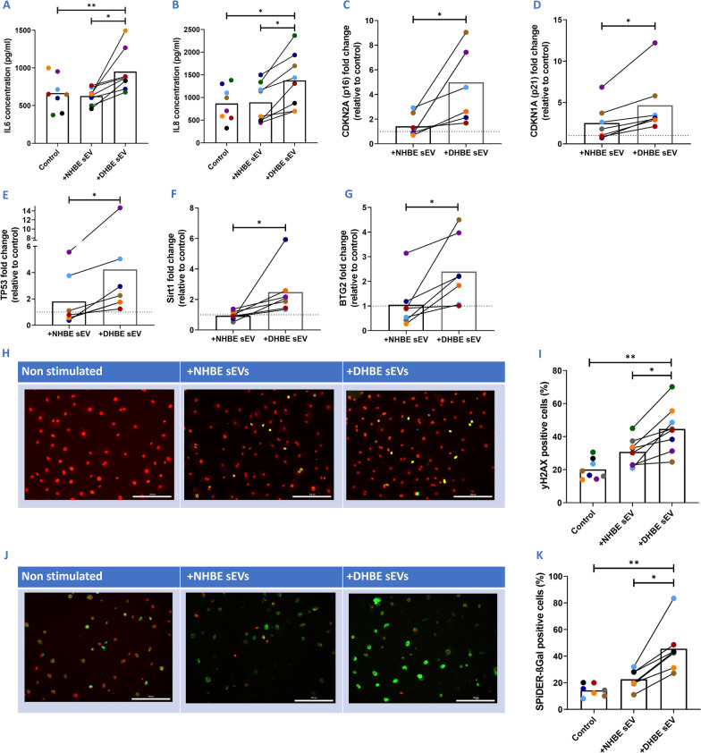
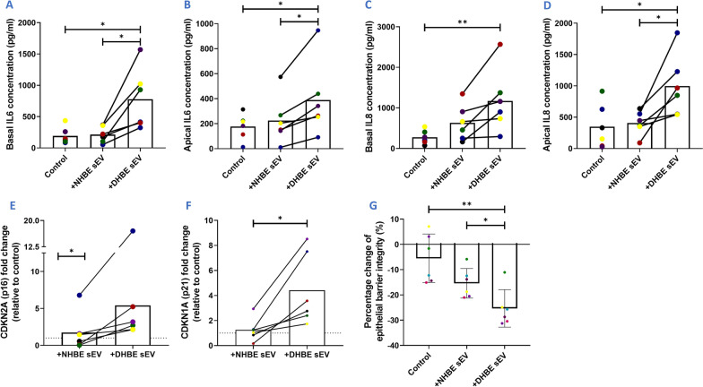
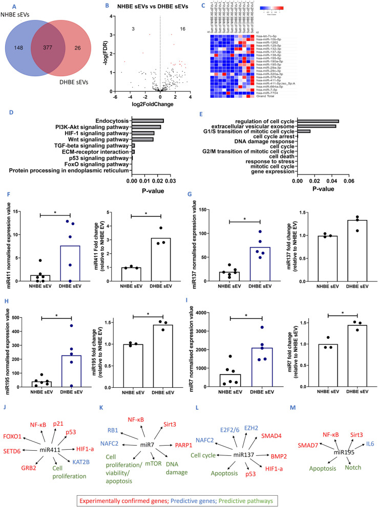
Increased sEV release from DHBEs compared to NHBEs (n = 4; p < 0.05) was detected by nanoparticle tracking analysis. NHBEs co-cultured with DHBE-derived sEVs for 72 h expressed higher levels of SA-β-Gal and γH2AX protein, p16 and p21 RNA and increased secretion of IL6 and IL8 proteins (all n = 6-8; p < 0.05). sEVs were also co-cultured with healthy air-liquid interface (ALI) cultures and similar results were observed, with increases in p21 and p16 gene expression and IL6 and IL8 (basal and apical) secretion (n = 6; p < 0.05). Transepithelial electrical resistance (TEER) measurements, a reflection of epithelial barrier integrity, were decreased upon the addition of DHBE-derived sEVs (n = 6; p < 0.05). smRNA-sequencing identified nineteen significantly differentially expressed miRNA in DHBE-derived sEVs compared to NHBE-derived sEVs, with candidate miRNAs validated by qPCR (all n = 5; p < 0.05). Four of these miRNAs were upregulated in NHBEs co-cultured with DHBE-derived sEVs and three in healthy ALI cultures co-cultured with DHBE-derived sEVs (n = 3-4; p < 0.05).
This data demonstrates that DHBE-derived sEVs transfer senescence to neighbouring healthy cells, promoting the disease state in IPF.
特发性肺纤维化(IPF)是一种影响全球 300 万人的慢性肺部疾病。衰老和小细胞外囊泡(sEVs)已被牵连到 IPF 的发病机制中,尽管 sEVs 如何促进疾病尚不清楚。在这里,我们对支气管上皮细胞中的 sEVs 进行了分析,并确定了小 RNA(smRNA)的含量。
收集条件培养基,并从正常人支气管上皮细胞(NHBEs)和特发性肺纤维化患者支气管上皮细胞(DHBEs)中分离 sEVs。
通过纳米颗粒跟踪分析检测到 DHBE 细胞比 NHBE 细胞释放的 sEV 明显增加(n=4;p<0.05)。NHBE 细胞与 DHBE 来源的 sEV 共培养 72 小时后,SA-β-Gal 和 γH2AX 蛋白、p16 和 p21 RNA 的表达水平更高,IL6 和 IL8 蛋白的分泌量也增加(所有 n=6-8;p<0.05)。sEV 还与健康的气液界面(ALI)培养物共培养,观察到类似的结果,p21 和 p16 基因表达以及 IL6 和 IL8(基底和顶端)分泌增加(n=6;p<0.05)。DHBE 来源的 sEV 加入后,上皮细胞屏障完整性的反映—跨上皮电阻(TEER)测量值降低(n=6;p<0.05)。DHBE 来源的 sEV 与 NHBE 来源的 sEV 相比,smRNA 测序确定了 19 个明显差异表达的 miRNA,通过 qPCR 验证了候选 miRNA(所有 n=5;p<0.05)。在与 DHBE 来源的 sEV 共培养的 NHBEs 中上调了 4 个 miRNA,在与 DHBE 来源的 sEV 共培养的健康 ALI 培养物中上调了 3 个 miRNA(n=3-4;p<0.05)。
这项数据表明,DHBE 来源的 sEV 将衰老转移到邻近的健康细胞,促进了 IPF 中的疾病状态。