Department of Orthopaedics and Trauma-Surgery, Medical University of Vienna, Währinger Gürtel 18-20, 1090, Vienna, Austria.
Michael Ogon Laboratory for Orthopaedic Research, Orthopaedic Hospital Vienna Speising, Speisinger Straße 109, 1130, Vienna, Austria.
Int Orthop. 2023 Apr;47(4):945-953. doi: 10.1007/s00264-023-05722-z. Epub 2023 Feb 17.
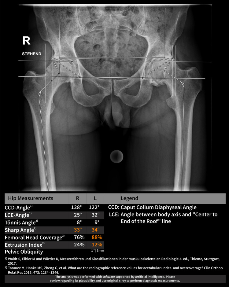
Despite advances of three-dimensional imaging pelvic radiographs remain the cornerstone in the evaluation of the hip joint. However, large inter- and intra-rater variabilities were reported due to subjective landmark setting. Artificial intelligence (AI)-powered software applications could improve the reproducibility of pelvic radiograph evaluation by providing standardized measurements. The aim of this study was to evaluate the reliability and agreement of a newly developed AI algorithm for the evaluation of pelvic radiographs.
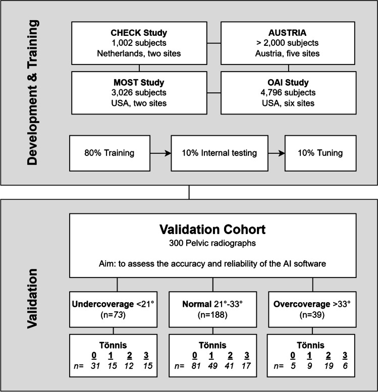
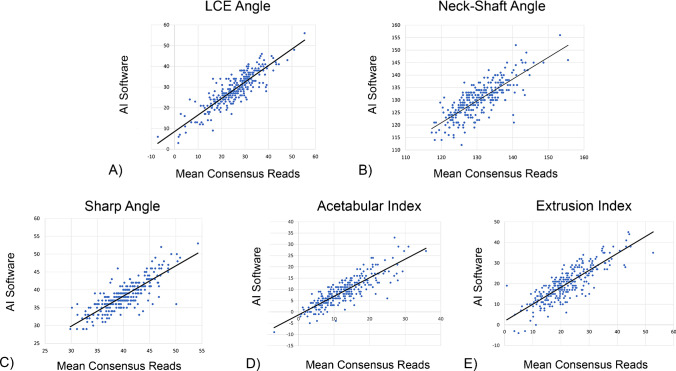
Three-hundred pelvic radiographs from 280 patients with different degrees of acetabular coverage and osteoarthritis (Tönnis Grade 0 to 3) were evaluated. Reliability and agreement between manual measurements and the outputs of the AI software were assessed for the lateral-center-edge (LCE) angle, neck-shaft angle, sharp angle, acetabular index, as well as the femoral head extrusion index.
The AI software provided reliable results in 94.3% (283/300). The ICC values ranged between 0.73 for the Acetabular Index to 0.80 for the LCE Angle. Agreement between readers and AI outputs, given by the standard error of measurement (SEM), was good for hips with normal coverage (LCE-SEM: 3.4°) and no osteoarthritis (LCE-SEM: 3.3°) and worse for hips with undercoverage (LCE-SEM: 5.2°) or severe osteoarthritis (LCE-SEM: 5.1°).
AI-powered applications are a reliable alternative to manual evaluation of pelvic radiographs. While being accurate for patients with normal acetabular coverage and mild signs of osteoarthritis, it needs improvement in the evaluation of patients with hip dysplasia and severe osteoarthritis.
尽管三维成像技术取得了进展,但骨盆 X 线片仍然是髋关节评估的基石。然而,由于主观地标设置,报道称存在较大的观察者内和观察者间变异性。人工智能(AI)驱动的软件应用程序可以通过提供标准化测量来提高骨盆 X 线片评估的可重复性。本研究旨在评估一种新开发的 AI 算法在评估骨盆 X 线片方面的可靠性和一致性。
评估了 280 名患者的 300 张骨盆 X 光片,这些患者的髋臼覆盖程度和骨关节炎程度不同(Tönnis 分级 0 至 3)。评估了手动测量值和 AI 软件输出值之间的侧向中心边缘(LCE)角、颈干角、锐度角、髋臼指数以及股骨头挤出指数的可靠性和一致性。
AI 软件提供了 94.3%(283/300)可靠的结果。ICC 值范围为髋臼指数为 0.73,LCE 角为 0.80。对于具有正常覆盖的髋关节(LCE-SEM:3.4°)和无骨关节炎的髋关节(LCE-SEM:3.3°),读者和 AI 输出之间的一致性由测量标准误差(SEM)给出,结果良好,而对于覆盖不足的髋关节(LCE-SEM:5.2°)或严重骨关节炎的髋关节(LCE-SEM:5.1°),则一致性较差。
AI 驱动的应用程序是手动评估骨盆 X 光片的可靠替代方法。虽然对于具有正常髋臼覆盖和轻度骨关节炎迹象的患者准确,但在评估髋关节发育不良和严重骨关节炎患者时需要改进。