Mental Health Service, Hospital Universitario Virgen del Rocío, Seville, Spain; Centro de Investigación Biomédica en Red de Salud Mental, Instituto de Salud Carlos III (CIBERSAM), Madrid, Spain; and Instituto de Biomedicina de Sevilla (IBiS)/HUVR/CSIC/Universidad de Sevilla, Seville, Spain.
Mental Health Service, Hospital Universitario Virgen del Rocío, Seville, Spain; Centro de Investigación Biomédica en Red de Salud Mental, Instituto de Salud Carlos III (CIBERSAM), Madrid, Spain; Instituto de Biomedicina de Sevilla (IBiS), Seville, Spain; and Department of Psychiatry, Universidad de Sevilla, Seville, Spain.
Br J Psychiatry. 2023 Jul;223(1):309-318. doi: 10.1192/bjp.2022.192.
Understanding the evolution of negative symptoms in first-episode psychosis (FEP) requires long-term longitudinal study designs that capture the progression of this condition and the associated brain changes.
To explore the factors underlying negative symptoms and their association with long-term abnormal brain trajectories.
We followed up 357 people with FEP over a 10-year period. Factor analyses were conducted to explore negative symptom dimensionality. Latent growth mixture modelling (LGMM) was used to identify the latent classes. Analysis of variance (ANOVA) was conducted to investigate developmental trajectories of cortical thickness. Finally, the resulting ANOVA maps were correlated with a wide set of regional molecular profiles derived from public databases.
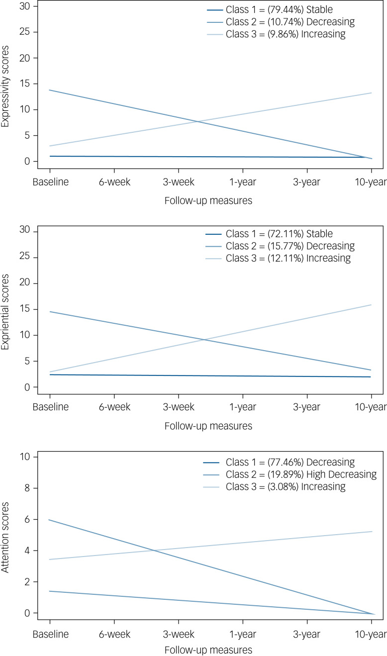
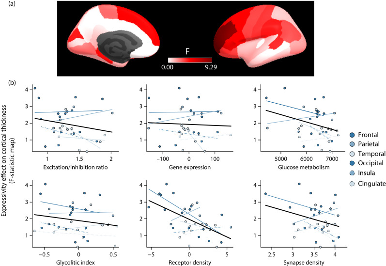
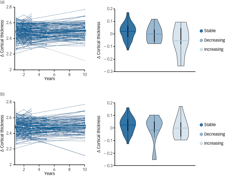
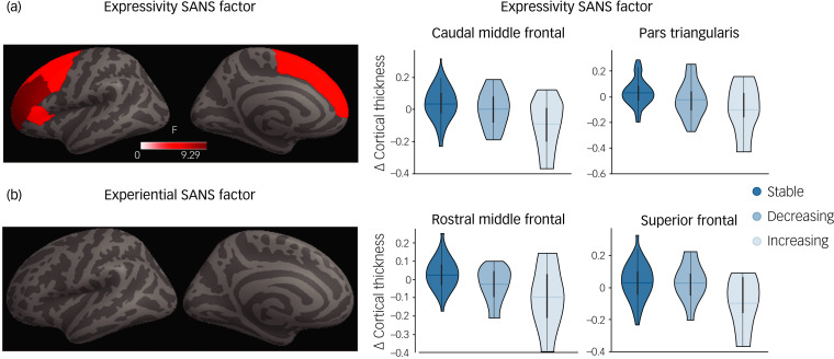
Three trajectories (stable, decreasing and increasing) were found in each of the three factors (expressivity, experiential and attention) identified by the factor analyses. Patients with an increasing trajectory in the expressivity factor showed cortical thinning in caudal middle frontal, pars triangularis, rostral middle frontal and superior frontal regions from the third to the tenth year after the onset of the psychotic disorder. The -statistic map of cortical thickness expressivity differences was associated with a receptor density map derived from positron emission tomography data.
Stable and decreasing were the most common trajectories. Additionally, cortical thickness abnormalities found at relatively late stages of FEP onset could be exploited as a biomarker of poor symptom outcome in the expressivity dimension. Finally, the brain areas with less density of receptors spatially overlap areas that discriminate the trajectories of the expressivity dimension.
了解首发精神病(FEP)患者阴性症状的演变需要长期的纵向研究设计,以捕捉该疾病的进展及其相关的大脑变化。
探讨阴性症状的潜在因素及其与长期异常大脑轨迹的关系。
我们对 357 名 FEP 患者进行了为期 10 年的随访。进行因子分析以探索阴性症状的维度。采用潜在增长混合模型(LGMM)来识别潜在的类别。方差分析(ANOVA)用于研究皮质厚度的发展轨迹。最后,将所得的 ANOVA 图谱与从公共数据库中获得的广泛的区域分子图谱进行相关分析。
在因子分析中确定的三个因子(表达性、体验性和注意力)中,每个因子都发现了三种轨迹(稳定、下降和增加)。在表达性因子中呈增加轨迹的患者,从精神病发病后的第三年到第十年,在尾侧中额、三角部、额前和额上回出现皮质变薄。皮质厚度表达差异的 -统计量图谱与正电子发射断层扫描数据衍生的受体密度图谱相关。
稳定和下降是最常见的轨迹。此外,在 FEP 发病相对较晚阶段发现的皮质厚度异常可作为表达维度不良症状结局的生物标志物。最后,受体密度较低的脑区与区分表达维度轨迹的脑区空间重叠。