Liu Jinrui, Xu Mengying, Ren Jialiang, Li Zhihao, Xi Lu, Chen Bing
School of Clinical Medicine, Ningxia Medical University, Yinchuan, China.
Department of Radiology, General Hospital of Ningxia Medical University, Yinchuan, China.
Front Oncol. 2023 Feb 3;12:1080580. doi: 10.3389/fonc.2022.1080580. eCollection 2022.
To assess the diagnostic value of predictive models based on synthetic magnetic resonance imaging (syMRI), multiplexed sensitivity encoding (MUSE) sequences, and Breast Imaging Reporting and Data System (BI-RADS) in the differentiation of benign and malignant breast lesions.
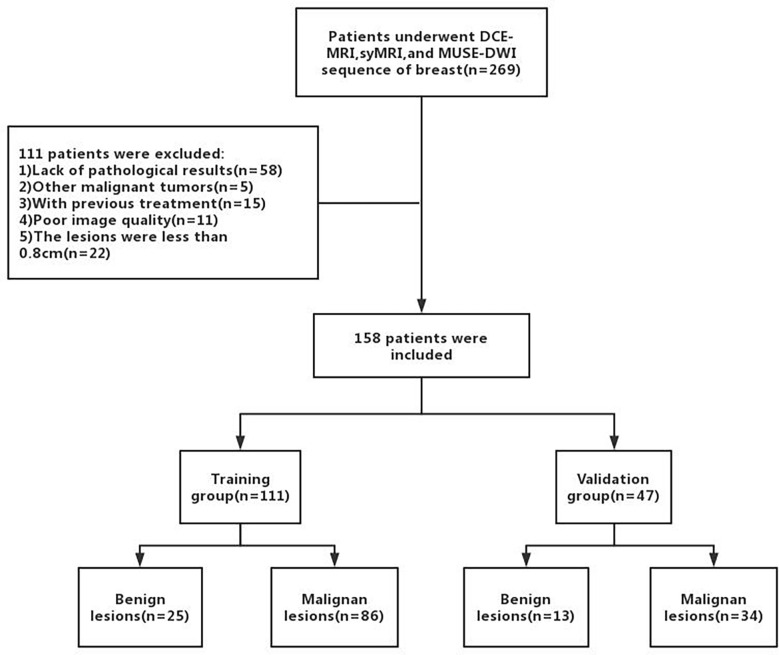
Clinical and MRI data of 158 patients with breast lesions who underwent dynamic contrast-enhanced MRI (DCE-MRI), syMRI, and MUSE sequences between September 2019 and December 2020 were retrospectively collected. The apparent diffusion coefficient (ADC) values of MUSE and quantitative relaxation parameters (longitudinal and transverse relaxation times [T1, T2], and proton density [PD] values) of syMRI were measured, and the parameter variation values and change in their ratios were calculated. The patients were randomly divided into training (n = 111) and validation (n = 47) groups at a ratio of 7:3. A nomogram was built based on univariate and multivariate logistic regression analyses in the training group and was verified in the validation group. The discriminatory and predictive capacities of the nomogram were assessed by the receiver operating characteristic curve and area under the curve (AUC). The AUC was compared by DeLong test.
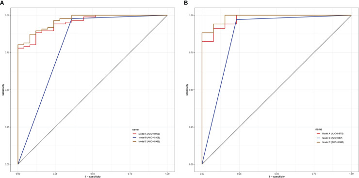
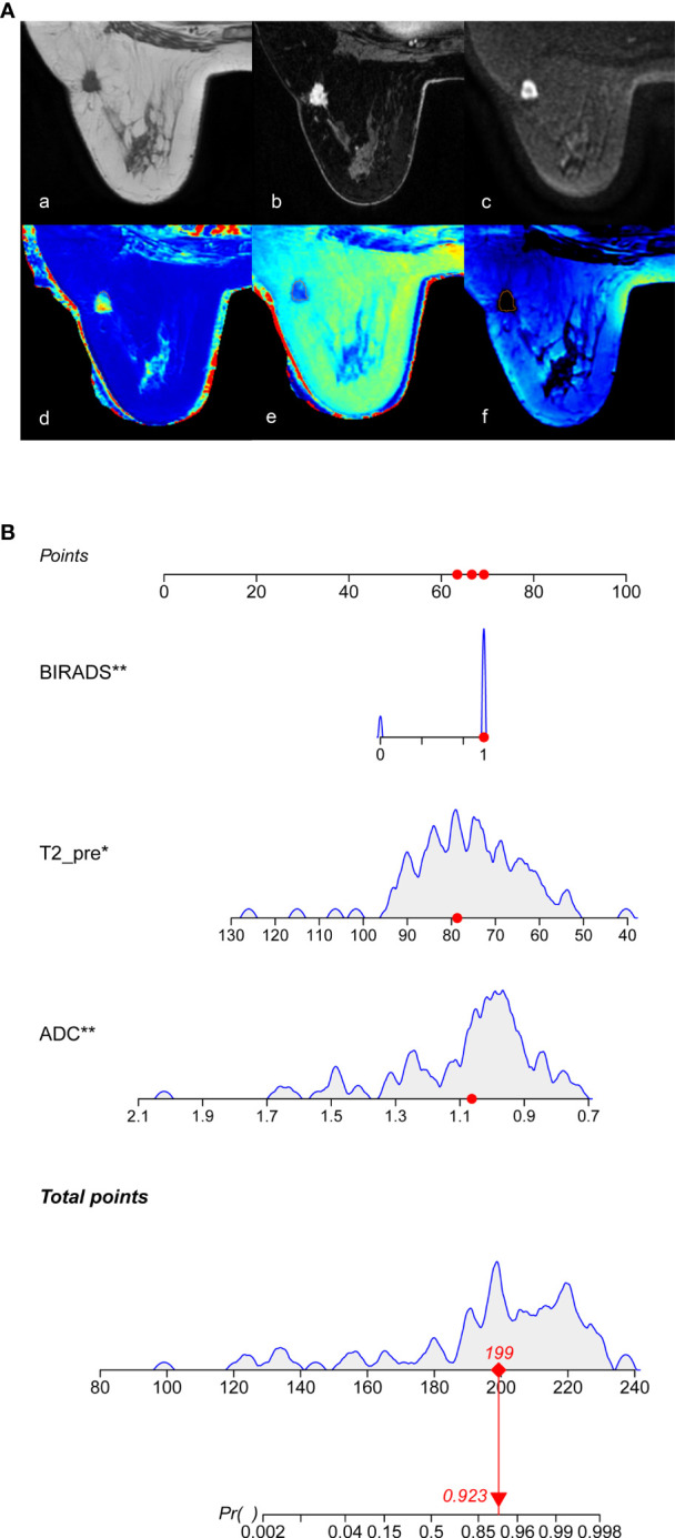
In the training group, univariate analysis showed that age, lesion diameter, menopausal status, ADC, T2, PD, PD, T2, and T2 were significantly different between benign and malignant breast lesions ( < 0.05). Multivariate logistic regression analysis showed that ADC and T2 were significant variables (all < 0.05) in breast cancer diagnosis. The quantitative model (model A: ADC, T2), BI-RADS model (model B), and multi-parameter model (model C: ADC, T2, BI-RADS) were established by combining the above independent variables, among which model C had the highest diagnostic performance, with AUC of 0.965 and 0.986 in the training and validation groups, respectively.
The prediction model established based on syMRI, MUSE sequence, and BI-RADS is helpful for clinical differentiation of breast tumors and provides more accurate information for individualized diagnosis.
评估基于合成磁共振成像(syMRI)、多通道敏感性编码(MUSE)序列以及乳腺影像报告和数据系统(BI-RADS)的预测模型在鉴别乳腺良恶性病变中的诊断价值。
回顾性收集2019年9月至2020年12月期间158例接受动态对比增强磁共振成像(DCE-MRI)、syMRI及MUSE序列检查的乳腺病变患者的临床和MRI数据。测量MUSE序列的表观扩散系数(ADC)值以及syMRI的定量弛豫参数(纵向和横向弛豫时间[T1、T2]以及质子密度[PD]值),并计算参数变化值及其比值变化。患者按7:3的比例随机分为训练组(n = 111)和验证组(n = 47)。在训练组中基于单因素和多因素逻辑回归分析构建列线图,并在验证组中进行验证。通过受试者操作特征曲线和曲线下面积(AUC)评估列线图的鉴别能力和预测能力。采用DeLong检验比较AUC。
在训练组中,单因素分析显示乳腺良恶性病变在年龄、病变直径、绝经状态、ADC、T2、PD、PD、T2和T2方面存在显著差异(<0.05)。多因素逻辑回归分析显示,ADC和T2是乳腺癌诊断中的显著变量(均<0.05)。通过组合上述自变量建立了定量模型(模型A:ADC、T2)、BI-RADS模型(模型B)和多参数模型(模型C:ADC、T2、BI-RADS),其中模型C的诊断性能最高,在训练组和验证组中的AUC分别为0.965和0.986。
基于syMRI、MUSE序列和BI-RADS建立的预测模型有助于乳腺肿瘤的临床鉴别,为个体化诊断提供更准确的信息。