Key Laboratory of Aging and Cancer Biology of Zhejiang Province, Department of Immunology and Pathogen Biology, School of Basic Medical Sciences, Hangzhou Normal University, Hangzhou, 311121, China.
State Key Laboratory for Diagnosis and Treatment of Infectious Diseases, The First Affiliated Hospital, College of Medicine, Zhejiang University, Hangzhou, 310003, China.
BMC Biol. 2023 Feb 24;21(1):42. doi: 10.1186/s12915-023-01542-0.
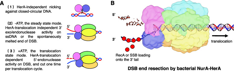
The nuclease NurA and the ATPase/translocase HerA play a vital role in repair of double-strand breaks (DSB) during the homologous recombination in archaea. A NurA-HerA complex is known to mediate DSB DNA end resection, leading to formation of a free 3' end used to search for the homologous sequence. Despite the structures of individual archaeal types of NurA and HerA having been reported, there is limited information regarding the molecular mechanisms underlying this process. Some bacteria also possess homologs of NurA and HerA; however, the bacterial type of this complex, as well as the detailed mechanisms underlying the joining of NurA-HerA in DSB DNA end resection, remains unclear.
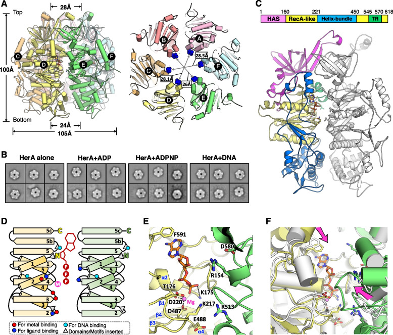
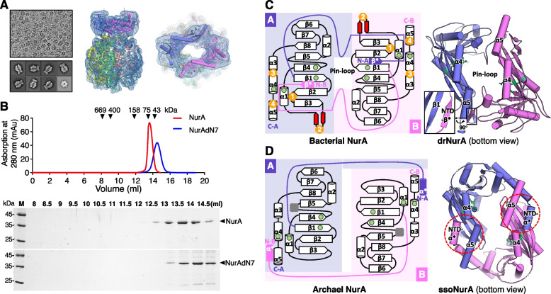
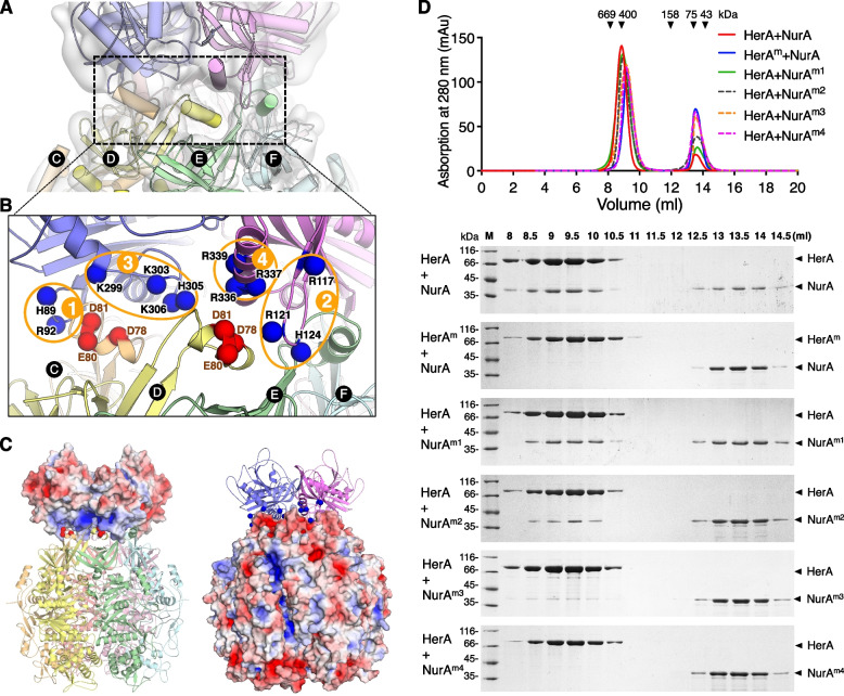
We report for the first time the crystal structures of Deinococcus radiodurans HerA (drHerA) in the nucleotide-free and ADP-binding modes. A D. radiodurans NurA-HerA complex structure was constructed according to a low-resolution cryo-electron microscopy map. We performed site-directed mutagenesis to map the drNurA-HerA interaction sites, suggesting that their interaction is mainly mediated by ionic links, in contrast to previously characterized archaeal NurA-HerA interactions. The key residues responsible for the DNA translocation activity, DNA unwinding activity, and catalytic activities of the drNurA-HerA complex were identified. A HerA/FtsK-specific translocation-related motif (TR motif) that guarantees the processivity of double-stranded DNA (dsDNA) translocation was identified. Moreover, a mechanism for the translocation-regulated resection of the 5' tail of broken dsDNA and the corresponding generation of a recombinogenic 3' single-stranded DNA tail by the drNurA-HerA complex was elucidated.
Our work provides new insights into the mechanism underlying bacterial NurA-HerA-mediated DSB DNA end resection, and the way this complex digests the 5' tail of a DNA duplex and provides long 3' free end for strand invasion in the bacterial homologous recombination process.
在古菌同源重组过程中,核酸内切酶 NurA 和 ATP 酶/转运酶 HerA 对于双链断裂(DSB)的修复起着至关重要的作用。已知 NurA-HerA 复合物介导 DSB DNA 末端切除,导致形成游离的 3' 末端,用于搜索同源序列。尽管已经报道了单个古菌类型的 NurA 和 HerA 的结构,但关于该过程的分子机制的信息有限。一些细菌也拥有 NurA 和 HerA 的同源物;然而,该复合物的细菌类型以及 NurA-HerA 在 DSB DNA 末端切除中连接的详细机制尚不清楚。
我们首次报道了 Deinococcus radiodurans HerA(drHerA)在核苷酸游离和 ADP 结合两种形式下的晶体结构。根据低分辨率冷冻电镜图谱构建了 D. radiodurans NurA-HerA 复合物结构。我们进行了定点突变以绘制 drNurA-HerA 相互作用位点图谱,表明它们的相互作用主要通过离子键介导,与以前表征的古菌 NurA-HerA 相互作用不同。确定了负责 drNurA-HerA 复合物 DNA 易位活性、DNA 解旋活性和催化活性的关键残基。鉴定了一个 HerA/FtsK 特异性易位相关基序(TR 基序),该基序保证了双链 DNA(dsDNA)易位的连续性。此外,阐明了 drNurA-HerA 复合物对断裂 dsDNA 的 5' 尾的易位调控切除以及由此产生的重组性 3' 单链 DNA 尾的生成机制。
我们的工作为细菌 NurA-HerA 介导的 DSB DNA 末端切除的机制以及该复合物在细菌同源重组过程中消化 DNA 双链的 5' 尾并为链入侵提供长的 3' 游离末端提供了新的见解。