• 文献检索
  • 文档翻译
  • 深度研究
  • 学术资讯
  • Suppr Zotero 插件Zotero 插件
  • 邀请有礼
  • 套餐&价格
  • 历史记录
应用&插件
Suppr Zotero 插件Zotero 插件浏览器插件Mac 客户端Windows 客户端微信小程序
定价
高级版会员购买积分包购买API积分包
服务
文献检索文档翻译深度研究API 文档MCP 服务
关于我们
关于 Suppr公司介绍联系我们用户协议隐私条款
关注我们

Suppr 超能文献

核心技术专利:CN118964589B侵权必究
粤ICP备2023148730 号-1Suppr @ 2026

文献检索

告别复杂PubMed语法,用中文像聊天一样搜索,搜遍4000万医学文献。AI智能推荐,让科研检索更轻松。

立即免费搜索

文件翻译

保留排版,准确专业,支持PDF/Word/PPT等文件格式,支持 12+语言互译。

免费翻译文档

深度研究

AI帮你快速写综述,25分钟生成高质量综述,智能提取关键信息,辅助科研写作。

立即免费体验

心率显著降低可显著提高经胸多普勒超声心动图检查时记录冠状动脉血流速度的可行性。

Heart Rate Lowering Significantly Increases Feasibility in Doppler Recording Blood Flow Velocity in Coronaries during Transthoracic Doppler Echocardiography.

作者信息

Caiati Carlo, Pollice Paolo, Lepera Mario Erminio

机构信息

Institute of Cardiovascular Disease, Department of Interdisciplinary Medicine, University of Bari "Aldo Moro", 70124 Bari, Italy.

出版信息

Diagnostics (Basel). 2023 Feb 10;13(4):670. doi: 10.3390/diagnostics13040670.

DOI:10.3390/diagnostics13040670
PMID:36832158
原文链接:https://pmc.ncbi.nlm.nih.gov/articles/PMC9955744/
Abstract

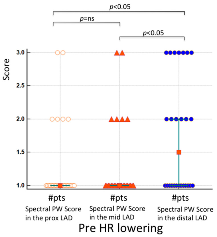
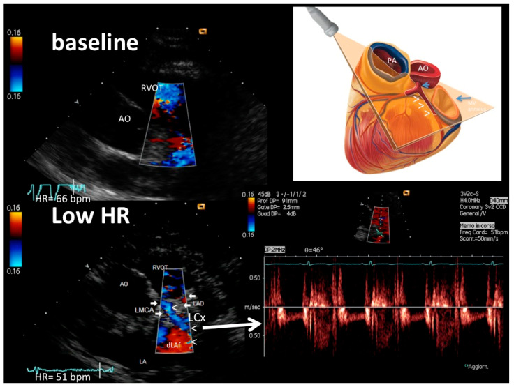
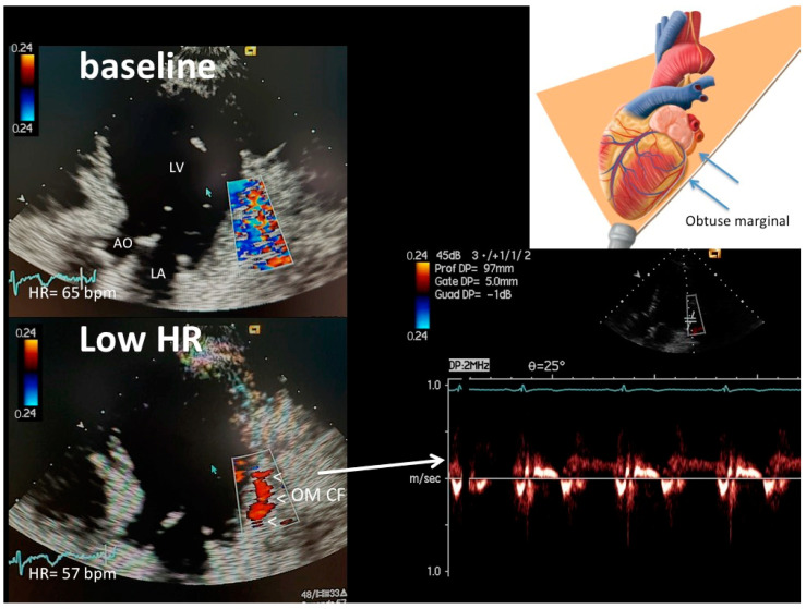
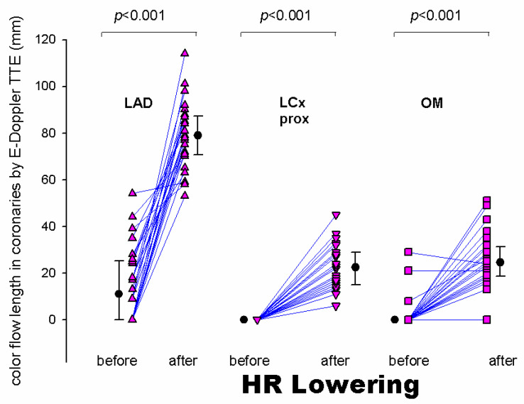
: Coronary blood flow Doppler recording by Transthoracic Doppler in convergent mode (E-Doppler TTE) might be further improved by lowering heart rate (HRL) down to <60 bpm, since low HR < 60 b/m causes a disproportional lengthening of the diastole, so the coronaries are still for a longer time, very much improving the Doppler signal/noise ratio. : A group of 26 patients underwent E-Doppler TTE before and after HR lowering in four branches of the coronary tree, namely, the left main (LMCA); left anterior descending (LAD), subdivided into three segments: proximal, mid and distal; proximal left circumflex (LCx); and obtuse marginal (OM). Color and PW coronary Doppler signal was judged by two expert observers as undetectable (SCORE 1), weak or with clutter artifacts (SCORE 2), or well delineated (SCORE 3). In addition, local accelerated stenotic flow (AsF) was measured in the LAD before and after HRL. : Beta-blockers significantly decreased the mean HR from 76 ± 5 to 57 ± 6 bpm ( < 0.001). Before HRL, the Doppler quality was very poor in the proximal and mid-LAD segments (median score value = 1 in both), while in the distal LAD, it was significantly better but still suboptimal (median score value = 1.5, = 0.009 vs. proximal and mid-LAD score). After HRL, blood flow Doppler recording in the three LAD segments was strikingly improved (median score value = 3, 3 and 3, = ns), so the effect of HRL was more efficacious in the two more proximal LAD segments. In 10 patients undergoing coronary angiography (CA), no AsF as expression of transtenotic velocity was detected at baseline. After HRL, thanks to the better quality and length of color flow, ASF was detected in five patients while in five others, it was not in perfect agreement with CA (Spearman correlation coefficient = 1, < 0.01). The color flow in the proximal LCx and OM was extremely poor at baseline (color flow length 0 and 0, median (interquartile range) mm, respectively) and improved considerably after HRL (color flow length 23 [13.5] and 25 [12.0] mm, respectively, < 0.001). : HRL greatly improved the success rate of blood flow Doppler recording in coronaries, not only in the LAD, but also in the LCx. Therefore, AsF for stenosis detection and coronary flow reserve assessment can have wider clinical applications. However, further studies with larger samples are needed to confirm these results.

摘要

经胸多普勒在收敛模式下(E-Doppler TTE)进行冠状动脉血流多普勒记录,通过将心率(HRL)降低至<60次/分钟可能会进一步改善,因为低心率<60次/分钟会导致舒张期不成比例地延长,因此冠状动脉处于舒张期的时间更长,这极大地提高了多普勒信号/噪声比。一组26例患者在心率降低前后,对冠状动脉树的四个分支进行了E-Doppler TTE检查,这四个分支分别是左主干(LMCA);左前降支(LAD),细分为三段:近端、中段和远端;近端左旋支(LCx);钝缘支(OM)。两名专家观察者将彩色和脉冲波冠状动脉多普勒信号判定为不可检测(评分1)、微弱或有杂波伪像(评分2)或清晰可辨(评分3)。此外,在心率降低前后测量了LAD处的局部加速狭窄血流(AsF)。β受体阻滞剂使平均心率从76±5次/分钟显著降低至57±6次/分钟(<0.001)。在心率降低前,LAD近端和中段的多普勒质量非常差(两者的中位数评分值均为1),而在LAD远端,质量明显更好但仍未达到最佳(中位数评分值为1.5,与近端和中段LAD评分相比,P=0.009)。心率降低后,LAD三个节段的血流多普勒记录显著改善(中位数评分值均为3,P=无显著差异),因此心率降低对LAD近端两个节段的效果更显著。在10例行冠状动脉造影(CA)的患者中,基线时未检测到作为跨狭窄速度表达的AsF。心率降低后,由于彩色血流质量更好、长度更长,5例患者检测到了AsF,而另外5例患者检测到的结果与CA不完全一致(Spearman相关系数=1,P<0.01)。LCx近端和OM的彩色血流在基线时极差(彩色血流长度分别为0和0,中位数(四分位间距)mm),心率降低后显著改善(彩色血流长度分别为23[13.5]和25[12.0]mm,P<0.001)。心率降低极大地提高了冠状动脉血流多普勒记录的成功率,不仅在LAD,在LCx也如此。因此,用于狭窄检测和冠状动脉血流储备评估的AsF可具有更广泛的临床应用。然而,需要更大样本量的进一步研究来证实这些结果。

https://cdn.ncbi.nlm.nih.gov/pmc/blobs/d5ff/9955744/6022c3f1a4eb/diagnostics-13-00670-g005.jpg
https://cdn.ncbi.nlm.nih.gov/pmc/blobs/d5ff/9955744/4665169531ab/diagnostics-13-00670-g001.jpg
https://cdn.ncbi.nlm.nih.gov/pmc/blobs/d5ff/9955744/dc2f1dc663e5/diagnostics-13-00670-g002.jpg
https://cdn.ncbi.nlm.nih.gov/pmc/blobs/d5ff/9955744/41991fb2a4f0/diagnostics-13-00670-g003.jpg
https://cdn.ncbi.nlm.nih.gov/pmc/blobs/d5ff/9955744/896b650f6020/diagnostics-13-00670-g004.jpg
https://cdn.ncbi.nlm.nih.gov/pmc/blobs/d5ff/9955744/6022c3f1a4eb/diagnostics-13-00670-g005.jpg

相似文献

1
Heart Rate Lowering Significantly Increases Feasibility in Doppler Recording Blood Flow Velocity in Coronaries during Transthoracic Doppler Echocardiography.心率显著降低可显著提高经胸多普勒超声心动图检查时记录冠状动脉血流速度的可行性。
Diagnostics (Basel). 2023 Feb 10;13(4):670. doi: 10.3390/diagnostics13040670.
2
A new noninvasive method for assessing mild coronary atherosclerosis: transthoracic convergent color Doppler after heart rate reduction. Validation vs. intracoronary ultrasound.一种评估轻度冠状动脉粥样硬化的新无创方法:心率降低后的经胸汇聚式彩色多普勒。与冠状动脉内超声的对比验证。
Coron Artery Dis. 2020 Sep;31(6):500-511. doi: 10.1097/MCA.0000000000000873.
3
Coronary Flow and Reserve by Enhanced Transthoracic Doppler Trumps Coronary Anatomy by Computed Tomography in Assessing Coronary Artery Stenosis.在评估冠状动脉狭窄方面,经胸多普勒增强技术测定的冠状动脉血流及储备优于计算机断层扫描测定的冠状动脉解剖结构。
Diagnostics (Basel). 2021 Feb 5;11(2):245. doi: 10.3390/diagnostics11020245.
4
Contrast-enhanced transthoracic second harmonic echo Doppler with adenosine: a noninvasive, rapid and effective method for coronary flow reserve assessment.经胸二次谐波造影增强回声多普勒与腺苷联合应用:一种评估冠状动脉血流储备的无创、快速且有效的方法。
J Am Coll Cardiol. 1999 Jul;34(1):122-30. doi: 10.1016/s0735-1097(99)00164-3.
5
Detection of angiographically significant coronary obstruction using resting transthoracic coronary Doppler echocardiography.使用静息经胸冠状动脉多普勒超声心动图检测血管造影显示有意义的冠状动脉阻塞。
J Echocardiogr. 2018 Sep;16(3):139-146. doi: 10.1007/s12574-017-0366-0. Epub 2017 Dec 16.
6
Accelerated stenotic flow in the left anterior descending coronary artery explains the causes of impaired coronary flow reserve: an integrated transthoracic enhanced Doppler study.经胸增强多普勒综合研究:左前降支冠状动脉狭窄血流加速解释了冠状动脉血流储备受损的原因
Front Cardiovasc Med. 2023 Sep 8;10:1186983. doi: 10.3389/fcvm.2023.1186983. eCollection 2023.
7
Noninvasive assessment of significant left anterior descending coronary artery stenosis by coronary flow velocity reserve with transthoracic color Doppler echocardiography.经胸彩色多普勒超声心动图通过冠状动脉血流储备对左前降支冠状动脉严重狭窄进行无创评估。
Circulation. 1998 Apr 28;97(16):1557-62. doi: 10.1161/01.cir.97.16.1557.
8
Demonstration of recanalized left coronary artery after thrombolysis by transthoracic echocardiography.经胸超声心动图显示溶栓后左冠状动脉再通。
J Am Soc Echocardiogr. 2005 Jun;18(6):686-92. doi: 10.1016/j.echo.2004.08.047.
9
Assessment of the Severity of Left Anterior Descending Coronary Artery Stenoses by Enhanced Transthoracic Doppler Echocardiography: Validation of a Method Based on the Continuity Equation.经胸增强多普勒超声心动图评估左前降支冠状动脉狭窄程度:基于连续性方程的方法验证
Diagnostics (Basel). 2023 Jul 29;13(15):2526. doi: 10.3390/diagnostics13152526.
10
Measurement of coronary flow velocity reserve in the posterior descending coronary artery by contrast-enhanced transthoracic Doppler echocardiography.经胸对比增强多普勒超声心动图测量后降支冠状动脉血流储备速度
J Am Soc Echocardiogr. 2004 Jan;17(1):21-7. doi: 10.1016/j.echo.2003.09.001.

引用本文的文献

1
Comparative study of myocardial perfusion and coronary flow velocity reserve derived from adenosine triphosphate stress myocardial contrast echocardiography in coronary lesions with no/mild stenosis.三磷酸腺苷负荷心肌对比超声心动图评估无/轻度狭窄冠状动脉病变心肌灌注及冠状动脉血流储备的对比研究
Front Cardiovasc Med. 2024 Sep 23;11:1353736. doi: 10.3389/fcvm.2024.1353736. eCollection 2024.
2
Accelerated stenotic flow in the left anterior descending coronary artery explains the causes of impaired coronary flow reserve: an integrated transthoracic enhanced Doppler study.经胸增强多普勒综合研究:左前降支冠状动脉狭窄血流加速解释了冠状动脉血流储备受损的原因
Front Cardiovasc Med. 2023 Sep 8;10:1186983. doi: 10.3389/fcvm.2023.1186983. eCollection 2023.
3

本文引用的文献

1
Wellens' Syndrome from COVID-19 Infection Assessed by Enhanced Transthoracic Coronary Echo Doppler: A Case Report.经增强经胸冠状动脉回声多普勒评估的新型冠状病毒肺炎感染所致Wellens综合征:一例报告
Diagnostics (Basel). 2022 Mar 25;12(4):804. doi: 10.3390/diagnostics12040804.
2
Doppler Echocardiography Assessment of Coronary Microvascular Function in Patients With Angina and No Obstructive Coronary Artery Disease.心绞痛且无阻塞性冠状动脉疾病患者冠状动脉微血管功能的多普勒超声心动图评估
Front Cardiovasc Med. 2021 Oct 29;8:723542. doi: 10.3389/fcvm.2021.723542. eCollection 2021.
3
A new noninvasive method for assessing mild coronary atherosclerosis: transthoracic convergent color Doppler after heart rate reduction. Validation vs. intracoronary ultrasound.
Assessment of the Severity of Left Anterior Descending Coronary Artery Stenoses by Enhanced Transthoracic Doppler Echocardiography: Validation of a Method Based on the Continuity Equation.经胸增强多普勒超声心动图评估左前降支冠状动脉狭窄程度:基于连续性方程的方法验证
Diagnostics (Basel). 2023 Jul 29;13(15):2526. doi: 10.3390/diagnostics13152526.
4
Free Radicals and Obesity-Related Chronic Inflammation Contrasted by Antioxidants: A New Perspective in Coronary Artery Disease.抗氧化剂对自由基与肥胖相关慢性炎症的作用对比:冠状动脉疾病的新视角
Metabolites. 2023 May 31;13(6):712. doi: 10.3390/metabo13060712.
一种评估轻度冠状动脉粥样硬化的新无创方法:心率降低后的经胸汇聚式彩色多普勒。与冠状动脉内超声的对比验证。
Coron Artery Dis. 2020 Sep;31(6):500-511. doi: 10.1097/MCA.0000000000000873.
4
Coronary Physiology Beyond Coronary Flow Reserve in Microvascular Angina: JACC State-of-the-Art Review.微血管性心绞痛中心肌血流储备以外的冠状动脉生理学:JACC 现状评价。
J Am Coll Cardiol. 2018 Nov 27;72(21):2642-2662. doi: 10.1016/j.jacc.2018.07.106.
5
Efficacy of Ivabradine versus β-Blockers for Heart Rate Reduction during Computed Tomography Coronary Angiography: A Meta-Analysis of Randomized Controlled Trials.伊伐布雷定与β受体阻滞剂在计算机断层扫描冠状动脉造影期间降低心率的疗效比较:一项随机对照试验的荟萃分析
Cardiology. 2016;135(3):133-140. doi: 10.1159/000447236. Epub 2016 Jun 22.
6
Transthoracic Doppler for detection of stenoses in the three main coronary arteries by use of stenotic to prestenotic velocity ratio and aliased coronary flow.经胸多普勒超声检测三主冠状动脉狭窄:狭窄前-狭窄后流速比值和冠状动脉血流频移。
Eur Heart J Cardiovasc Imaging. 2015 Dec;16(12):1323-30. doi: 10.1093/ehjci/jev158. Epub 2015 Jun 25.
7
Coronary flow velocity reserve in the three main coronary arteries assessed with transthoracic Doppler: a comparative study with quantitative coronary angiography.经胸多普勒评估的三支主要冠状动脉的冠状动脉血流储备:与定量冠状动脉造影的对比研究。
J Am Soc Echocardiogr. 2011 Jul;24(7):758-67. doi: 10.1016/j.echo.2011.03.010. Epub 2011 Apr 23.
8
Detection, location, and severity assessment of left anterior descending coronary artery stenoses by means of contrast-enhanced transthoracic harmonic echo Doppler.通过对比增强经胸谐波回声多普勒检测、定位和评估左前降支冠状动脉狭窄的程度
Eur Heart J. 2009 Jul;30(14):1797-806. doi: 10.1093/eurheartj/ehp163. Epub 2009 May 9.
9
Non-invasive assessment of coronary flow and coronary flow reserve by transthoracic Doppler echocardiography: a magic tool for the real world.经胸多普勒超声心动图对冠状动脉血流及冠状动脉血流储备的无创评估:现实世界中的神奇工具。
Eur J Echocardiogr. 2008 Jul;9(4):449-57. doi: 10.1093/ejechocard/jen004. Epub 2008 Feb 19.
10
Distal left circumflex coronary artery flow reserve recorded by transthoracic Doppler echocardiography: a comparison with Doppler-wire.经胸多普勒超声心动图记录的左回旋支远端冠状动脉血流储备:与多普勒导丝的比较。
Cardiovasc Ultrasound. 2007 Jun 16;5:22. doi: 10.1186/1476-7120-5-22.