• 文献检索
  • 文档翻译
  • 深度研究
  • 学术资讯
  • Suppr Zotero 插件Zotero 插件
  • 邀请有礼
  • 套餐&价格
  • 历史记录
应用&插件
Suppr Zotero 插件Zotero 插件浏览器插件Mac 客户端Windows 客户端微信小程序
定价
高级版会员购买积分包购买API积分包
服务
文献检索文档翻译深度研究API 文档MCP 服务
关于我们
关于 Suppr公司介绍联系我们用户协议隐私条款
关注我们

Suppr 超能文献

核心技术专利:CN118964589B侵权必究
粤ICP备2023148730 号-1Suppr @ 2026

文献检索

告别复杂PubMed语法,用中文像聊天一样搜索,搜遍4000万医学文献。AI智能推荐,让科研检索更轻松。

立即免费搜索

文件翻译

保留排版,准确专业,支持PDF/Word/PPT等文件格式,支持 12+语言互译。

免费翻译文档

深度研究

AI帮你快速写综述,25分钟生成高质量综述,智能提取关键信息,辅助科研写作。

立即免费体验

转录因子 AKNA 在 T 细胞活化中的关键作用:一种综合生物信息学方法。

Critical Role of the Transcription Factor AKNA in T-Cell Activation: An Integrative Bioinformatics Approach.

机构信息

Center for Research on Infectious Diseases, Instituto Nacional de Salud Pública, Cuernavaca 62100, Mexico.

Department of Biochemistry and Molecular Genetics, Feinberg School of Medicine, Northwestern University, Chicago, IL 60611, USA.

出版信息

Int J Mol Sci. 2023 Feb 20;24(4):4212. doi: 10.3390/ijms24044212.

DOI:10.3390/ijms24044212
PMID:36835622
原文链接:https://pmc.ncbi.nlm.nih.gov/articles/PMC9965657/
Abstract

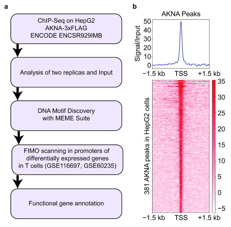
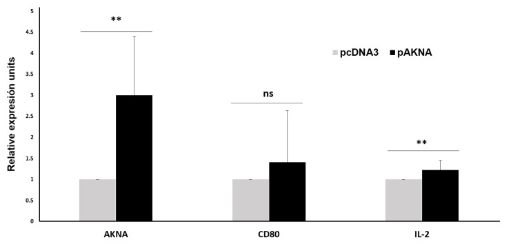
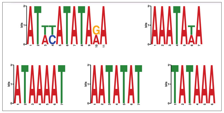
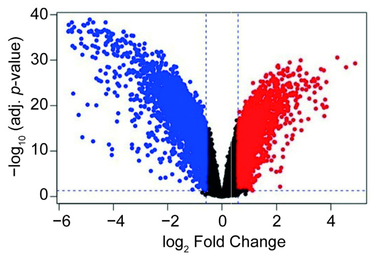
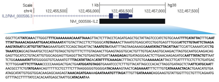
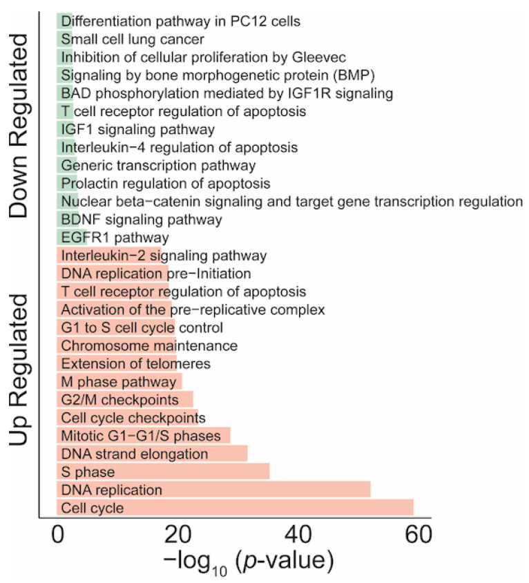
The human gene encodes an AT-hook transcription factor, the expression of which is involved in various cellular processes. The goal of this study was to identify potential AKNA binding sites in genes that participate in T-cell activation and validate selected genes. Here we analyzed ChIP-seq and microarray assays to determine AKNA-binding motifs and the cellular process altered by AKNA in T-cell lymphocytes. In addition, we performed a validation analysis by RT-qPCR to assess AKNA's role in promoting and expression. We found five AT-rich motifs that are potential candidates as AKNA response elements. We identified these AT-rich motifs in promoter regions of more than a thousand genes in activated T-cells, and demonstrated that AKNA induces the expression of genes involved in helper T-cell activation, such as . The genomic enrichment and prediction of AT-rich motif analyses demonstrated that AKNA is a transcription factor that can potentially modulate gene expression by recognizing AT-rich motifs in a plethora of genes that are involved in different molecular pathways and processes. Among the cellular processes activated by AT-rich genes, we found inflammatory pathways potentially regulated by AKNA, suggesting AKNA is acting as a master regulator during T-cell activation.

摘要

该人类基因编码一个 AT 钩转录因子,其表达参与各种细胞过程。本研究的目的是鉴定参与 T 细胞激活的基因中潜在的 AKNA 结合位点,并验证选定的基因。在这里,我们分析了 ChIP-seq 和微阵列分析,以确定 AKNA 在 T 细胞淋巴细胞中结合的基序和改变的细胞过程。此外,我们通过 RT-qPCR 进行了验证分析,以评估 AKNA 在促进和表达中的作用。我们发现了五个可能作为 AKNA 反应元件的富含 AT 的基序。我们在激活的 T 细胞中的超过一千个基因的启动子区域中鉴定出这些富含 AT 的基序,并证明 AKNA 诱导参与辅助 T 细胞激活的基因的表达,如。基因组富集和富含 AT 的基序分析预测表明,AKNA 是一种转录因子,可以通过识别参与不同分子途径和过程的众多基因中的富含 AT 的基序,潜在地调节基因表达。在富含 AT 的基因激活的细胞过程中,我们发现了潜在受 AKNA 调节的炎症途径,表明 AKNA 在 T 细胞激活过程中充当主调控因子。

https://cdn.ncbi.nlm.nih.gov/pmc/blobs/ee2f/9965657/4344eb0fb9d3/ijms-24-04212-g006.jpg
https://cdn.ncbi.nlm.nih.gov/pmc/blobs/ee2f/9965657/12251e9f9842/ijms-24-04212-g001.jpg
https://cdn.ncbi.nlm.nih.gov/pmc/blobs/ee2f/9965657/89ae74ecdea4/ijms-24-04212-g002.jpg
https://cdn.ncbi.nlm.nih.gov/pmc/blobs/ee2f/9965657/1e07992c1b45/ijms-24-04212-g003.jpg
https://cdn.ncbi.nlm.nih.gov/pmc/blobs/ee2f/9965657/509800f817bd/ijms-24-04212-g004.jpg
https://cdn.ncbi.nlm.nih.gov/pmc/blobs/ee2f/9965657/e5107ad3184c/ijms-24-04212-g005.jpg
https://cdn.ncbi.nlm.nih.gov/pmc/blobs/ee2f/9965657/4344eb0fb9d3/ijms-24-04212-g006.jpg

相似文献

1
Critical Role of the Transcription Factor AKNA in T-Cell Activation: An Integrative Bioinformatics Approach.转录因子 AKNA 在 T 细胞活化中的关键作用:一种综合生物信息学方法。
Int J Mol Sci. 2023 Feb 20;24(4):4212. doi: 10.3390/ijms24044212.
2
PKA/CREB and NF-κB pathway regulates AKNA transcription: A novel insight into T-2 toxin-induced inflammation and GH deficiency in GH3 cells.蛋白激酶A/环磷腺苷效应元件结合蛋白与核因子κB信号通路调控AKNA转录:对T-2毒素诱导GH3细胞炎症和生长激素缺乏的新见解
Toxicology. 2017 Dec 1;392:81-95. doi: 10.1016/j.tox.2017.10.013. Epub 2017 Oct 25.
3
Regulation of CD40 and CD40 ligand by the AT-hook transcription factor AKNA.AT钩转录因子AKNA对CD40和CD40配体的调控
Nature. 2001 Mar 15;410(6826):383-7. doi: 10.1038/35066602.
4
Is a Potential Prognostic Biomarker in Gastric Cancer and Function as a Tumor Suppressor by Modulating EMT-Related Pathways.在胃癌中是一种潜在的预后生物标志物,并通过调节 EMT 相关途径发挥肿瘤抑制作用。
Biomed Res Int. 2020 May 13;2020:6726759. doi: 10.1155/2020/6726759. eCollection 2020.
5
Coordinate activation of inflammatory gene networks, alveolar destruction and neonatal death in AKNA deficient mice.AKNA 缺陷小鼠中炎症基因网络的协调激活、肺泡破坏和新生仔鼠死亡。
Cell Res. 2011 Nov;21(11):1564-77. doi: 10.1038/cr.2011.84. Epub 2011 May 24.
6
Functional Role of AKNA: A Scoping Review.AKNA 的功能作用:范围综述。
Biomolecules. 2021 Nov 17;11(11):1709. doi: 10.3390/biom11111709.
7
The human AKNA gene expresses multiple transcripts and protein isoforms as a result of alternative promoter usage, splicing, and polyadenylation.由于启动子的选择性使用、剪接和聚腺苷酸化,人类AKNA基因表达多种转录本和蛋白质异构体。
DNA Cell Biol. 2005 May;24(5):325-38. doi: 10.1089/dna.2005.24.325.
8
A Homozygous Frameshift Variant Is Associated with Microcephaly in a Pakistani Family.一个巴基斯坦家族的小头畸形与纯合移码变异有关。
Genes (Basel). 2021 Sep 24;12(10):1494. doi: 10.3390/genes12101494.
9
Cervical cancer-associated promoter polymorphism affects akna expression levels.宫颈癌相关启动子多态性影响 akna 表达水平。
Genes Immun. 2015 Jan-Feb;16(1):43-53. doi: 10.1038/gene.2014.60. Epub 2014 Nov 6.
10
The association of AKNA gene polymorphisms with knee osteoarthritis suggests the relevance of this immune response regulator in the disease genetic susceptibility.AKNA基因多态性与膝骨关节炎的关联表明这种免疫反应调节因子在疾病遗传易感性中的相关性。
Mol Biol Rep. 2018 Apr;45(2):151-161. doi: 10.1007/s11033-018-4148-1. Epub 2018 Jan 24.

引用本文的文献

1
Binding to the Other Side: The AT-Hook DNA-Binding Domain Allows Nuclear Factors to Exploit the DNA Minor Groove.与另一侧结合:AT 钩 DNA 结合结构域使核因子能够利用 DNA 小沟。
Int J Mol Sci. 2024 Aug 14;25(16):8863. doi: 10.3390/ijms25168863.

本文引用的文献

1
T lymphocytes induce human cancer cells derived from solid malignant tumors to secrete galectin-9 which facilitates immunosuppression in cooperation with other immune checkpoint proteins.T 淋巴细胞诱导源自实体恶性肿瘤的人类癌细胞分泌半乳糖凝集素-9,该分子与其他免疫检查点蛋白协同作用促进免疫抑制。
J Immunother Cancer. 2023 Jan;11(1). doi: 10.1136/jitc-2022-005714.
2
Functional Role of AKNA: A Scoping Review.AKNA 的功能作用:范围综述。
Biomolecules. 2021 Nov 17;11(11):1709. doi: 10.3390/biom11111709.
3
Investigation of the Impact from IL-2, IL-7, and IL-15 on the Growth and Signaling of Activated CD4 T Cells.
探讨白细胞介素 2(IL-2)、白细胞介素 7(IL-7)和白细胞介素 15(IL-15)对活化 CD4 T 细胞生长和信号转导的影响。
Int J Mol Sci. 2020 Oct 22;21(21):7814. doi: 10.3390/ijms21217814.
4
Targeted Activation of T Cells with IL-2-Coupled Nanoparticles.靶向激活 T 细胞的 IL-2 偶联纳米颗粒。
Cells. 2020 Sep 9;9(9):2063. doi: 10.3390/cells9092063.
5
Regulation of T Helper Cell Fate by TCR Signal Strength.T 辅助细胞命运的 TCR 信号强度调节。
Front Immunol. 2020 May 19;11:624. doi: 10.3389/fimmu.2020.00624. eCollection 2020.
6
HMGA Proteins in Hematological Malignancies.血液系统恶性肿瘤中的HMGA蛋白
Cancers (Basel). 2020 Jun 3;12(6):1456. doi: 10.3390/cancers12061456.
7
Roles of PD-1/PD-L1 Pathway: Signaling, Cancer, and Beyond.PD-1/PD-L1 通路的作用:信号转导、癌症及其他。
Adv Exp Med Biol. 2020;1248:33-59. doi: 10.1007/978-981-15-3266-5_3.
8
PD-1 and PD-L1 correlated gene expression profiles and their association with clinical outcomes of breast cancer.PD-1和PD-L1相关基因表达谱及其与乳腺癌临床结局的关联
Cancer Cell Int. 2019 Sep 9;19:233. doi: 10.1186/s12935-019-0955-2. eCollection 2019.
9
The Galaxy platform for accessible, reproducible and collaborative biomedical analyses: 2018 update.Galaxy 平台:用于可访问、可重复和协作的生物医学分析:2018 年更新。
Nucleic Acids Res. 2018 Jul 2;46(W1):W537-W544. doi: 10.1093/nar/gky379.
10
Lymphocyte Activation in Health and Disease.健康与疾病中的淋巴细胞激活
Crit Rev Immunol. 2017;37(2-6):439-462. doi: 10.1615/CritRevImmunol.v37.i2-6.120.