Aman Martin, Schulte Matthias, Li Yu, Thomas Benjamin, Daeschler Simeon, Mayrhofer-Schmid Maximilian, Kneser Ulrich, Harhaus Leila, Boecker Arne
Department of Hand, Plastic and Reconstructive Surgery, Burn Center, BG Trauma Center Ludwigshafen, Department of Hand- and Plastic Surgery, University of Heidelberg, Ludwig-Guttmann-Str. 13, 67071 Ludwigshafen, Germany.
J Clin Med. 2023 Feb 7;12(4):1306. doi: 10.3390/jcm12041306.
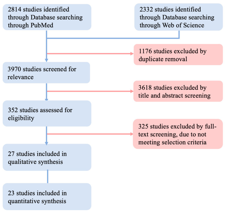
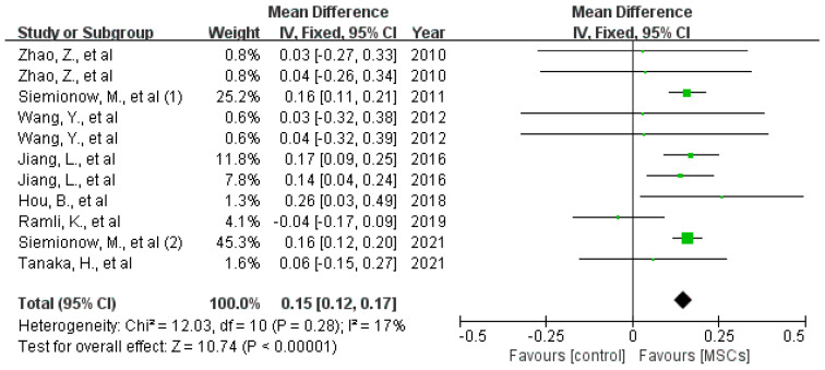
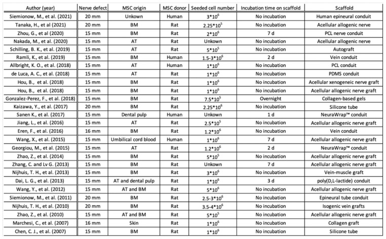
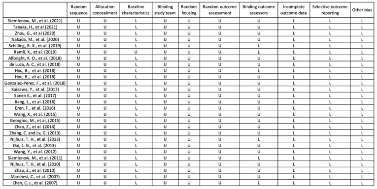
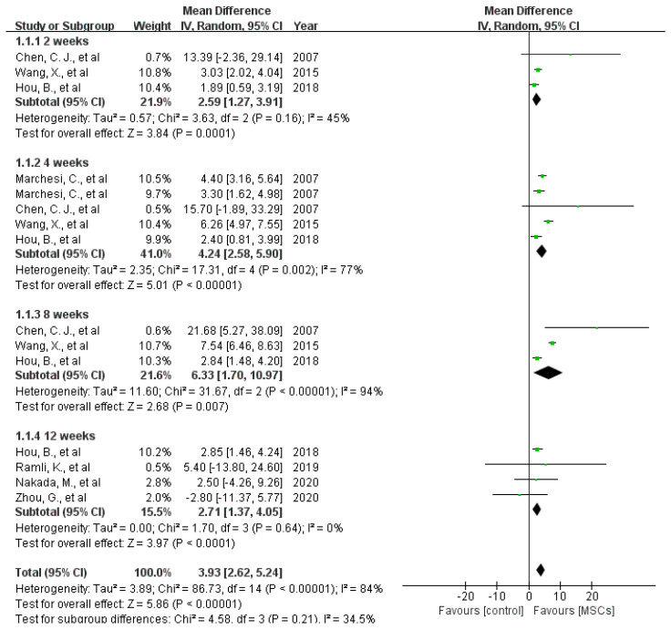
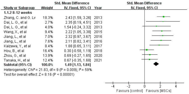
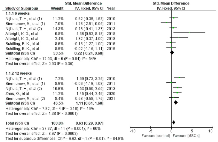
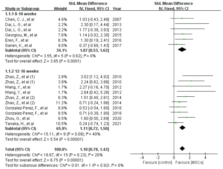
Critically sized nerve defects cause devastating life-long disabilities and require interposition for reconstruction. Additional local application of mesenchymal stem cells (MSCs) is considered promising to enhance peripheral nerve regeneration. To better understand the role of MSCs in peripheral nerve reconstruction, we performed a systematic review and meta-analysis of the effects of MSCs on critically sized segment nerve defects in preclinical studies. 5146 articles were screened following PRISMA guidelines using PubMed and Web of Science. A total of 27 preclinical studies (n = 722 rats) were included in the meta-analysis. The mean difference or the standardized mean difference with 95% confidence intervals for motor function, conduction velocity, and histomorphological parameters of nerve regeneration, as well as the degree of muscle atrophy, was compared in rats with critically sized defects and autologous nerve reconstruction treated with or without MSCs. The co-transplantation of MSCs increased the sciatic functional index (3.93, 95% CI 2.62 to 5.24, < 0.00001) and nerve conduction velocity recovery (1.49, 95% CI 1.13 to 1.84, = 0.009), decreased the atrophy of targeted muscles (gastrocnemius: 0.63, 95% CI 0.29 to 0.97 = 0.004; triceps surae: 0.08, 95% CI 0.06 to 0.10 = 0.71), and promoted the regeneration of injured axons (axon number: 1.10, 95% CI 0.78 to 1.42, < 0.00001; myelin sheath thickness: 0.15, 95% CI 0.12 to 0.17, = 0.28). Reconstruction of critically sized peripheral nerve defects is often hindered by impaired postoperative regeneration, especially in defects that require an autologous nerve graft. This meta-analysis indicates that additional application of MSC can enhance postoperative peripheral nerve regeneration in rats. Based on the promising results in vivo experiments, further studies are needed to demonstrate potential clinical benefits.
临界尺寸的神经缺损会导致严重的终身残疾,需要进行神经移植来重建。额外局部应用间充质干细胞(MSCs)被认为有望促进周围神经再生。为了更好地理解MSCs在周围神经重建中的作用,我们对临床前研究中MSCs对临界尺寸节段性神经缺损的影响进行了系统评价和荟萃分析。按照PRISMA指南,使用PubMed和Web of Science筛选了5146篇文章。荟萃分析共纳入27项临床前研究(n = 722只大鼠)。比较了在有临界尺寸缺损并接受或未接受MSCs治疗的自体神经重建的大鼠中,运动功能、传导速度、神经再生的组织形态学参数以及肌肉萎缩程度的平均差或95%置信区间的标准化平均差。MSCs的联合移植增加了坐骨神经功能指数(3.93,95%CI 2.62至5.24,<0.00001)和神经传导速度恢复(1.49,95%CI 1.13至1.84,=0.009),减少了目标肌肉的萎缩(腓肠肌:0.63,95%CI 0.29至0.97,=0.004;小腿三头肌:0.08,95%CI 0.06至0.10,=0.71),并促进了损伤轴突的再生(轴突数量:1.10,95%CI 0.78至1.42,<0.00001;髓鞘厚度:0.15,95%CI 0.12至0.17,=0.28)。临界尺寸周围神经缺损的重建常常因术后再生受损而受阻,尤其是在需要自体神经移植的缺损中。这项荟萃分析表明,额外应用MSCs可以增强大鼠术后周围神经再生。基于体内实验的良好结果,需要进一步研究以证明其潜在的临床益处。