Department of Orthopedic Surgery, the First Affiliated Hospital of Zhengzhou University, NO.1 Jianshe East Road, Zhengzhou, China.
Department of Orthopedic Surgery, the Honghui Hospital of Xi'an, No. 76 Nanguo road, Nan Xiaomen, Xi'an, 710054, China.
BMC Musculoskelet Disord. 2023 Feb 27;24(1):151. doi: 10.1186/s12891-023-06245-9.
3D printing technology has become a research hotspot in the field of scientific research because of its personalized customization, maneuverability and the ability to achieve multiple material fabrications. The focus of this study is to use 3D printing technology to customize personalized poly L-lactic acid (PLLA) porous screws in orthopedic plants and to explore its effect on tendon-bone healing after anterior cruciate ligament (ACL) reconstruction.
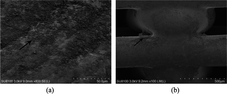
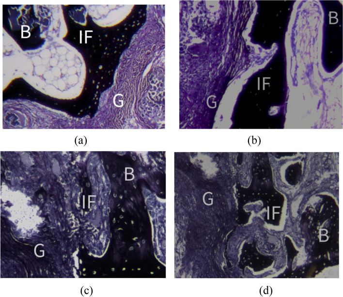
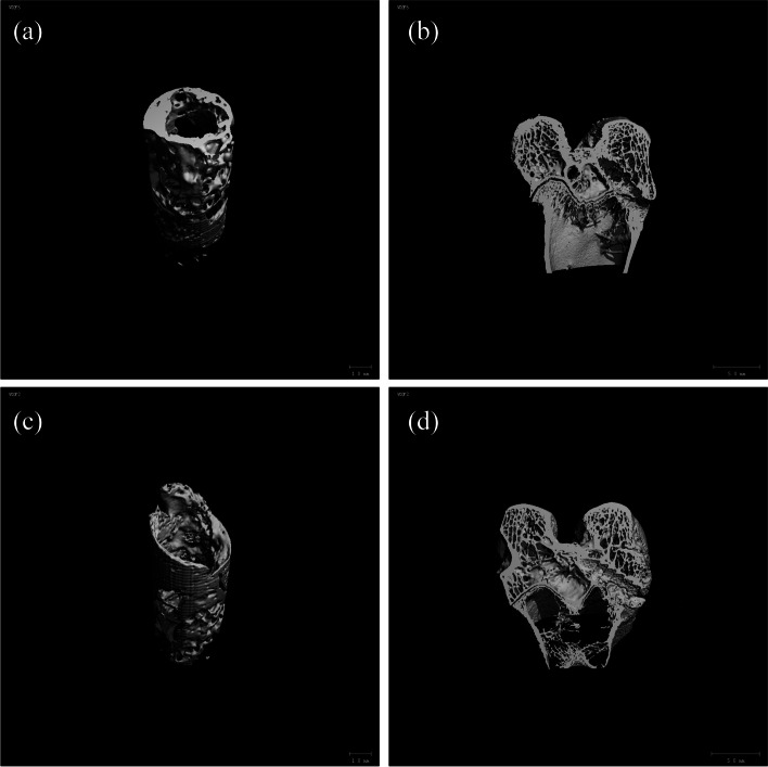
Preparation of PLLA porous screws with good orthogonal pore structure by 3D printer. The hydroxyapatite (HA) was adsorbed on porous screws by electrostatic layer-by-layer self-assembly (ELSA) technology, and PLLA-HA porous screws were prepared. The surface and spatial morphology of the modified screws were observed by scanning electron microscopy (SEM). The porosity of porous screw was measured by liquid displacement method. Thirty New Zealand male white rabbits were divided into two groups according to simple randomization. Autologous tendon was used for right ACL reconstruction, and porous screws were inserted into the femoral tunnel to fix the transplanted tendon. PLLA group was fixed with porous screws, PLLA-HA group was fixed with HA modified porous screws. At 6 weeks and 12 weeks after surgery, 5 animals in each group were sacrificed randomly for histological examination. The remaining 5 animals in each group underwent Micro-CT and biomechanical tests.
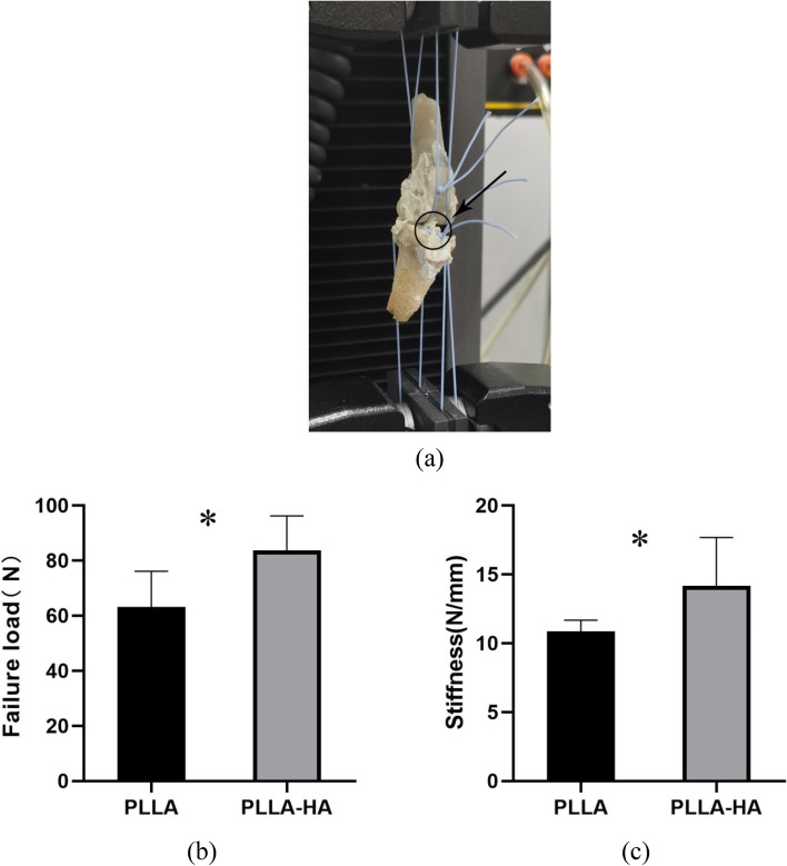
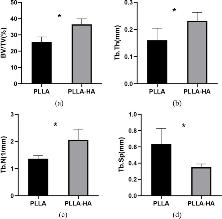
The pores of PLLA porous screws prepared by 3D printer were uniformly distributed and connected with each other, which meet the experimental requirements. HA was evenly distributed in the porous screw by ELSA technique. Histology showed that compared with PLLA group, mature bone trabeculae were integrated with grafted tendons in PLLA-HA group. Micro-CT showed that the bone formation index of PLLA-HA group was better than that of PLLA group. The new bone was uniformly distributed in the bone tunnel along the screw channel. Biomechanical experiments showed that the failure load and stiffness of PLLA-HA group were significantly higher than those of PLLA group.
The 3D printed PLLA porous screw modified by HA can not only fix the grafted tendons, but also increase the inductivity of bone, promote bone growth in the bone tunnel and promote bone integration at the tendon-bone interface. The PLLA-HA porous screw is likely to be used in clinic in the future.
3D 打印技术因其个性化定制、操作性强以及能够实现多种材料制造而成为科研领域的研究热点。本研究的重点是使用 3D 打印技术定制个性化聚左旋乳酸(PLLA)多孔螺钉,用于骨科植入物,并探讨其在前交叉韧带(ACL)重建后对腱骨愈合的影响。
使用 3D 打印机制备具有良好正交孔结构的 PLLA 多孔螺钉。通过静电层层自组装(ELSA)技术将羟基磷灰石(HA)吸附在多孔螺钉上,制备 PLLA-HA 多孔螺钉。通过扫描电子显微镜(SEM)观察改性螺钉的表面和空间形态。通过液体置换法测量多孔螺钉的孔隙率。30 只新西兰雄性白兔按简单随机分组法分为两组。自体肌腱用于右侧 ACL 重建,将多孔螺钉插入股骨隧道固定移植肌腱。PLLA 组用多孔螺钉固定,PLLA-HA 组用 HA 改性多孔螺钉固定。术后 6 周和 12 周,每组随机处死 5 只动物进行组织学检查。每组其余 5 只动物进行 Micro-CT 和生物力学测试。
3D 打印机制备的 PLLA 多孔螺钉的孔均匀分布并相互连通,满足实验要求。ELSA 技术使 HA 均匀分布在多孔螺钉中。组织学显示,与 PLLA 组相比,PLLA-HA 组成熟的骨小梁与移植物肌腱融合。Micro-CT 显示,PLLA-HA 组的骨形成指数优于 PLLA 组。新骨沿螺钉通道均匀分布在骨隧道内。生物力学实验表明,PLLA-HA 组的失效载荷和刚度明显高于 PLLA 组。
HA 改性的 3D 打印 PLLA 多孔螺钉不仅可以固定移植物肌腱,还可以增加骨的诱导性,促进骨隧道内骨生长,促进腱骨界面的骨整合。PLLA-HA 多孔螺钉有望在未来临床应用。