• 文献检索
  • 文档翻译
  • 深度研究
  • 学术资讯
  • Suppr Zotero 插件Zotero 插件
  • 邀请有礼
  • 套餐&价格
  • 历史记录
应用&插件
Suppr Zotero 插件Zotero 插件浏览器插件Mac 客户端Windows 客户端微信小程序
定价
高级版会员购买积分包购买API积分包
服务
文献检索文档翻译深度研究API 文档MCP 服务
关于我们
关于 Suppr公司介绍联系我们用户协议隐私条款
关注我们

Suppr 超能文献

核心技术专利:CN118964589B侵权必究
粤ICP备2023148730 号-1Suppr @ 2026

文献检索

告别复杂PubMed语法,用中文像聊天一样搜索,搜遍4000万医学文献。AI智能推荐,让科研检索更轻松。

立即免费搜索

文件翻译

保留排版,准确专业,支持PDF/Word/PPT等文件格式,支持 12+语言互译。

免费翻译文档

深度研究

AI帮你快速写综述,25分钟生成高质量综述,智能提取关键信息,辅助科研写作。

立即免费体验

采用两端改性羟基磷灰石的丝胶原支架重建兔前交叉韧带:组织学和生物力学研究。

Anterior cruciate ligament reconstruction in a rabbit model using a silk-collagen scaffold modified by hydroxyapatite at both ends: a histological and biomechanical study.

机构信息

Department of Orthopedic Surgery, The First Affiliated Hospital of Zhengzhou University, NO.1 Jianshe East Road, Zhengzhou, 450001, China.

Henan University of Chinese Medicine, NO.156 Jinshui East Road, Zhengzhou, 450001, China.

出版信息

J Orthop Surg Res. 2021 Feb 16;16(1):139. doi: 10.1186/s13018-021-02281-0.

DOI:10.1186/s13018-021-02281-0
PMID:33593365
原文链接:https://pmc.ncbi.nlm.nih.gov/articles/PMC7885370/
Abstract

BACKGROUND

To investigate osteointegration at the graft-bone interface and the prevention of osteoarthritis after anterior cruciate ligament (ACL) reconstruction using a silk-collagen scaffold with both ends modified by hydroxyapatite (HA) in a rabbit model.

METHODS

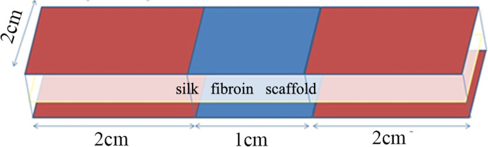
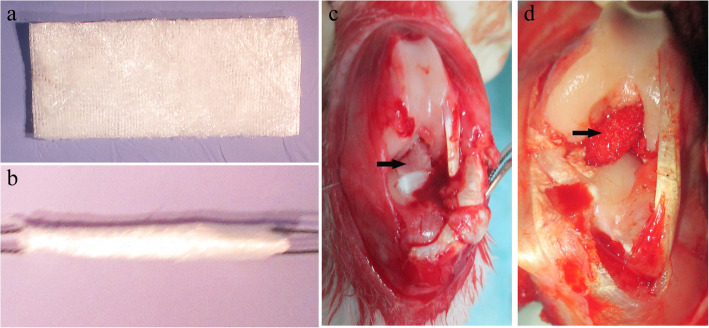
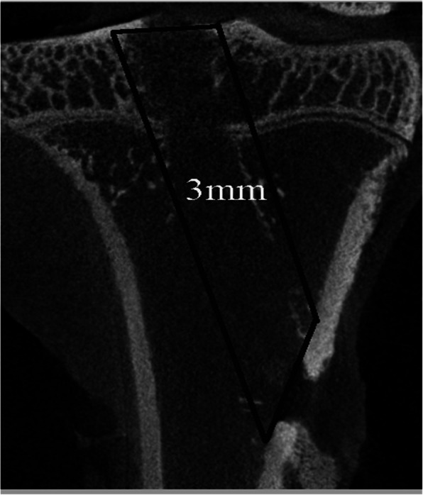
The HA/silk-collagen scaffold was fabricated using a degummed, knitted silk scaffold, collagen I matrix, and simulated body fluid (SBF). The HA/silk-collagen scaffold was rolled up to make a graft for replacing the native ACL in the experimental group (HA group), and the silk-collagen scaffold was used in the control (S group). All specimens were harvested at 16 weeks postoperatively to evaluate graft-bone healing and osteoarthritis prevention.

RESULTS

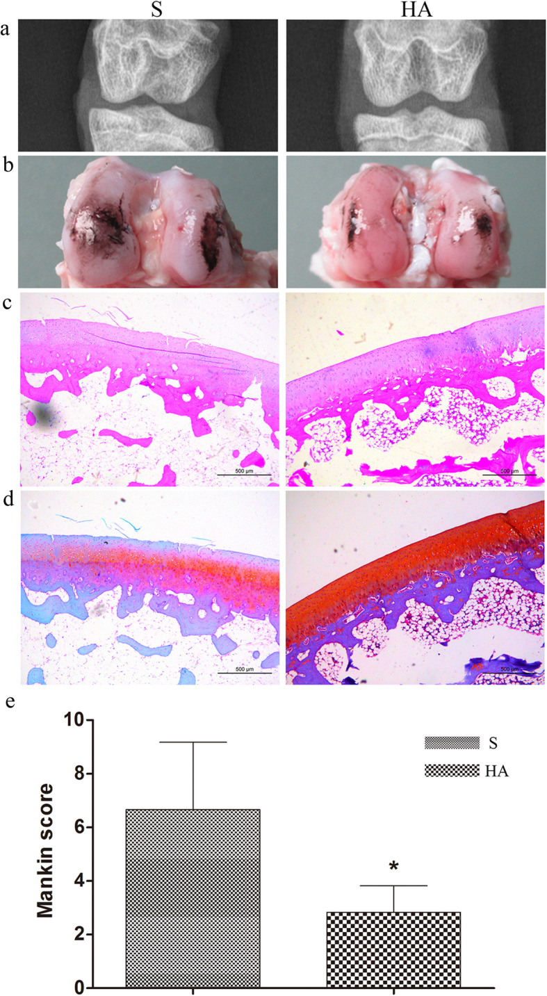
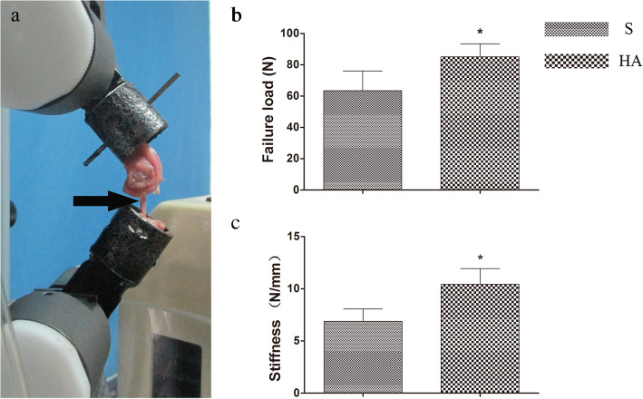
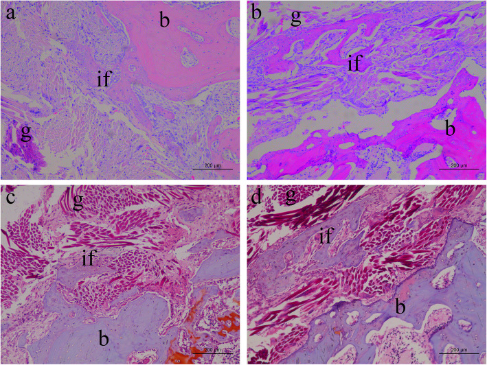
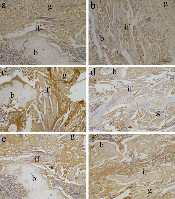
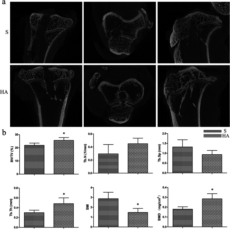
Histological staining revealed the massive formation of more mature bone at the tendon-bone interface, and immunohistochemistry staining revealed more collagen I and osteocalcin deposition in the HA group than in the S group. Higher signals indicating more bone mineral formation were detected in the HA group than in the S group, which was consistent with the results of biomechanical testing. Better osteoarthritis prevention was also observed in the HA group, indicating a more stable knee joint in the HA group than in the S group.

CONCLUSION

The HA/silk-collagen scaffold promotes osteointegration at the tendon-bone interface after ACL reconstruction and has great potential for clinical applications.

摘要

背景

为了在兔模型中研究 ACL 重建后使用两端经羟基磷灰石(HA)修饰的丝胶原支架的移植物-骨界面的骨整合和预防骨关节炎。

方法

使用脱胶、针织丝支架、I 型胶原基质和模拟体液(SBF)制备 HA/丝胶原支架。将 HA/丝胶原支架卷起制成替代天然 ACL 的移植物用于实验组(HA 组),而丝胶原支架用于对照组(S 组)。所有标本均在术后 16 周收获,以评估移植物-骨愈合和预防骨关节炎。

结果

组织学染色显示在肌腱-骨界面大量形成更成熟的骨,免疫组织化学染色显示在 HA 组中比在 S 组中沉积更多的 I 型胶原和骨钙素。在 HA 组中检测到更多的骨矿物质形成的信号,表明更多的骨形成,这与生物力学测试的结果一致。在 HA 组中也观察到更好的骨关节炎预防,表明 HA 组的膝关节比 S 组更稳定。

结论

HA/丝胶原支架促进 ACL 重建后肌腱-骨界面的骨整合,具有很大的临床应用潜力。

https://cdn.ncbi.nlm.nih.gov/pmc/blobs/fea8/7885370/8c787344fbfc/13018_2021_2281_Fig8_HTML.jpg
https://cdn.ncbi.nlm.nih.gov/pmc/blobs/fea8/7885370/349456e9cbba/13018_2021_2281_Fig1_HTML.jpg
https://cdn.ncbi.nlm.nih.gov/pmc/blobs/fea8/7885370/52f05baee64d/13018_2021_2281_Fig2_HTML.jpg
https://cdn.ncbi.nlm.nih.gov/pmc/blobs/fea8/7885370/751d7dc84970/13018_2021_2281_Fig3_HTML.jpg
https://cdn.ncbi.nlm.nih.gov/pmc/blobs/fea8/7885370/641be9db10cb/13018_2021_2281_Fig4_HTML.jpg
https://cdn.ncbi.nlm.nih.gov/pmc/blobs/fea8/7885370/da81b6fd6158/13018_2021_2281_Fig5_HTML.jpg
https://cdn.ncbi.nlm.nih.gov/pmc/blobs/fea8/7885370/4a2e71653b3f/13018_2021_2281_Fig6_HTML.jpg
https://cdn.ncbi.nlm.nih.gov/pmc/blobs/fea8/7885370/92f5b6dcad8d/13018_2021_2281_Fig7_HTML.jpg
https://cdn.ncbi.nlm.nih.gov/pmc/blobs/fea8/7885370/8c787344fbfc/13018_2021_2281_Fig8_HTML.jpg

相似文献

1
Anterior cruciate ligament reconstruction in a rabbit model using a silk-collagen scaffold modified by hydroxyapatite at both ends: a histological and biomechanical study.采用两端改性羟基磷灰石的丝胶原支架重建兔前交叉韧带:组织学和生物力学研究。
J Orthop Surg Res. 2021 Feb 16;16(1):139. doi: 10.1186/s13018-021-02281-0.
2
Long-term effects of knitted silk-collagen sponge scaffold on anterior cruciate ligament reconstruction and osteoarthritis prevention. knitted 丝胶原蛋白海绵支架对前交叉韧带重建和预防骨关节炎的长期影响。
Biomaterials. 2014 Sep;35(28):8154-63. doi: 10.1016/j.biomaterials.2014.06.019. Epub 2014 Jun 25.
3
Anterior cruciate ligament reconstruction in a rabbit model using silk-collagen scaffold and comparison with autograft.在兔模型中使用丝素-胶原支架进行前交叉韧带重建并与自体移植进行比较。
PLoS One. 2015 May 4;10(5):e0125900. doi: 10.1371/journal.pone.0125900. eCollection 2015.
4
[A PRELIMINARY STUDY ON SMALL INTESTINAL SUBMUCOSA-SILK COMPOSITE SCAFFOLD TO RECONSTRUCT ANTERIOR CRUCIATE LIGAMENT].小肠黏膜下层-丝素复合支架重建前交叉韧带的初步研究
Zhongguo Xiu Fu Chong Jian Wai Ke Za Zhi. 2015 Dec;29(12):1534-40.
5
Osteointegration of a Novel Silk Fiber-Based ACL Scaffold by Formation of a Ligament-Bone Interface.新型丝纤维 ACL 支架的骨整合:韧带-骨界面的形成。
Am J Sports Med. 2019 Mar;47(3):620-627. doi: 10.1177/0363546518818792. Epub 2019 Jan 17.
6
Decellularized Versus Fresh-Frozen Allografts in Anterior Cruciate Ligament Reconstruction: An In Vitro Study in a Rabbit Model.去细胞异体移植物与新鲜冷冻异体移植物在前交叉韧带重建中的应用:兔模型的体外研究
Am J Sports Med. 2015 Aug;43(8):1924-34. doi: 10.1177/0363546515585314. Epub 2015 Jun 2.
7
Three-Dimensional Bio-Printed Scaffold Sleeves With Mesenchymal Stem Cells for Enhancement of Tendon-to-Bone Healing in Anterior Cruciate Ligament Reconstruction Using Soft-Tissue Tendon Graft.三维生物打印支架袖带联合间充质干细胞促进前交叉韧带重建中使用软组织肌腱移植物的腱骨愈合
Arthroscopy. 2018 Jan;34(1):166-179. doi: 10.1016/j.arthro.2017.04.016. Epub 2017 Jul 6.
8
Bone Mesenchymal Stem Cells Contribute to Ligament Regeneration and Graft-Bone Healing after Anterior Cruciate Ligament Reconstruction with Silk-Collagen Scaffold.骨间充质干细胞有助于前交叉韧带重建术后使用丝胶原支架的韧带再生和移植骨愈合。
Stem Cells Int. 2021 Apr 23;2021:6697969. doi: 10.1155/2021/6697969. eCollection 2021.
9
The hydroxyapatite modified 3D printed poly L-lactic acid porous screw in reconstruction of anterior cruciate ligament of rabbit knee joint: a histological and biomechanical study.羟基磷灰石改性 3D 打印聚左旋乳酸多孔螺钉重建兔膝关节前交叉韧带:组织学和生物力学研究。
BMC Musculoskelet Disord. 2023 Feb 27;24(1):151. doi: 10.1186/s12891-023-06245-9.
10
Use of a bioactive scaffold to stimulate anterior cruciate ligament healing also minimizes posttraumatic osteoarthritis after surgery.使用生物活性支架刺激前交叉韧带愈合,也可以最大限度地减少手术后的创伤性骨关节炎。
Am J Sports Med. 2013 Aug;41(8):1762-70. doi: 10.1177/0363546513483446. Epub 2013 Jul 15.

引用本文的文献

1
A comparative study of collagen morphology and joint strength in anterior cruciate ligament repair and reconstruction models in rabbits.兔前交叉韧带修复与重建模型中胶原蛋白形态与关节强度的比较研究
Vet World. 2025 May;18(5):1313-1321. doi: 10.14202/vetworld.2025.1313-1321. Epub 2025 May 25.
2
Application and optimization of bioengineering strategies in facilitating tendon-bone healing.生物工程策略在促进肌腱-骨愈合中的应用与优化
Biomed Eng Online. 2025 Apr 23;24(1):46. doi: 10.1186/s12938-025-01368-7.
3
Enhancing tendon-bone integration and healing with advanced multi-layer nanofiber-reinforced 3D scaffolds for acellular tendon complexes.

本文引用的文献

1
Strategies for Using Polydopamine to Induce Biomineralization of Hydroxyapatite on Implant Materials for Bone Tissue Engineering.用于骨组织工程的植入材料上诱导羟基磷灰石生物矿化的聚多巴胺使用策略。
Int J Mol Sci. 2020 Sep 7;21(18):6544. doi: 10.3390/ijms21186544.
2
Biomineralizaion of hydroxyapatite on polyethylene terephthalate artificial ligaments promotes graft-bone healing after anterior cruciate ligament reconstruction: An in vitro and in vivo study.聚对苯二甲酸乙二醇酯人工韧带表面羟基磷灰石仿生矿化促进前交叉韧带重建后移植物-骨愈合:一项体内外研究。
J Biomater Appl. 2020 Aug;35(2):193-204. doi: 10.1177/0885328220921530. Epub 2020 Apr 26.
3
使用先进的多层纳米纤维增强三维支架促进无细胞肌腱复合体的腱骨整合与愈合。
Mater Today Bio. 2024 May 22;26:101099. doi: 10.1016/j.mtbio.2024.101099. eCollection 2024 Jun.
4
Silk fibroin-based scaffolds for tissue engineering.用于组织工程的丝素蛋白基支架
Front Bioeng Biotechnol. 2024 Apr 25;12:1381838. doi: 10.3389/fbioe.2024.1381838. eCollection 2024.
5
Advances in the application of hydrogel-based scaffolds for tendon repair.基于水凝胶的支架在肌腱修复中的应用进展。
Genes Dis. 2023 Jul 7;11(4):101019. doi: 10.1016/j.gendis.2023.04.039. eCollection 2024 Jul.
6
Biomimetic Intrafibrillar Mineralization of Native Tendon for Soft-Hard Interface Integration by Infiltration of Amorphous Calcium Phosphate Precursors.通过非晶态磷酸钙前体的渗透实现天然肌腱的仿生纤维内矿化,用于软-硬界面整合。
Adv Sci (Weinh). 2023 Dec;10(34):e2304216. doi: 10.1002/advs.202304216. Epub 2023 Oct 23.
7
Could an Anterior Cruciate Ligament Be Tissue-Engineered from Silk?能否从丝中构建前交叉韧带?
Cells. 2023 Sep 25;12(19):2350. doi: 10.3390/cells12192350.
8
Strategies to promote tendon-bone healing after anterior cruciate ligament reconstruction: Present and future.前交叉韧带重建术后促进腱骨愈合的策略:现状与未来。
Front Bioeng Biotechnol. 2023 Mar 13;11:1104214. doi: 10.3389/fbioe.2023.1104214. eCollection 2023.
9
The hydroxyapatite modified 3D printed poly L-lactic acid porous screw in reconstruction of anterior cruciate ligament of rabbit knee joint: a histological and biomechanical study.羟基磷灰石改性 3D 打印聚左旋乳酸多孔螺钉重建兔膝关节前交叉韧带:组织学和生物力学研究。
BMC Musculoskelet Disord. 2023 Feb 27;24(1):151. doi: 10.1186/s12891-023-06245-9.
10
Novel hybrid biocomposites for tendon grafts: The addition of silk to polydioxanone and poly(lactide-co-caprolactone) enhances material properties, and biocompatibility.用于肌腱移植的新型混合生物复合材料:在聚二氧六环酮和聚(丙交酯-共-己内酯)中添加丝可增强材料性能和生物相容性。
Bioact Mater. 2023 Feb 10;25:291-306. doi: 10.1016/j.bioactmat.2023.02.003. eCollection 2023 Jul.
Hydroxyapatite/silver electrospun fibers for anti-infection and osteoinduction.
用于抗感染和骨诱导的羟基磷灰石/银电纺纤维
J Adv Res. 2019 Oct 9;21:91-102. doi: 10.1016/j.jare.2019.10.002. eCollection 2020 Jan.
4
Enhance the biocompatibility and osseointegration of polyethylene terephthalate ligament by plasma spraying with hydroxyapatite in vitro and in vivo.通过等离子喷涂法在体外和体内将羟基磷灰石喷涂到聚对苯二甲酸乙二醇酯韧带上,以提高其生物相容性和骨整合性。
Int J Nanomedicine. 2018 Jun 25;13:3609-3623. doi: 10.2147/IJN.S162466. eCollection 2018.
5
Cell Therapies in Tendon, Ligament, and Musculoskeletal System Repair.细胞疗法在肌腱、韧带及肌肉骨骼系统修复中的应用
Sports Med Arthrosc Rev. 2018 Jun;26(2):48-58. doi: 10.1097/JSA.0000000000000192.
6
A review of hydroxyapatite-based coating techniques: Sol-gel and electrochemical depositions on biocompatible metals.基于羟基磷灰石的涂层技术综述:生物相容性金属上的溶胶-凝胶和电化学沉积法
J Mech Behav Biomed Mater. 2016 Apr;57:95-108. doi: 10.1016/j.jmbbm.2015.11.031. Epub 2015 Dec 6.
7
Novel therapeutic core-shell hydrogel scaffolds with sequential delivery of cobalt and bone morphogenetic protein-2 for synergistic bone regeneration.具有钴和骨形态发生蛋白-2顺序递送功能的新型治疗性核壳水凝胶支架用于协同骨再生。
Acta Biomater. 2015 Sep;23:295-308. doi: 10.1016/j.actbio.2015.06.002. Epub 2015 Jun 6.
8
In silico Mechano-Chemical Model of Bone Healing for the Regeneration of Critical Defects: The Effect of BMP-2.用于严重骨缺损再生的骨愈合计算机机械化学模型:骨形态发生蛋白-2的作用
PLoS One. 2015 Jun 4;10(6):e0127722. doi: 10.1371/journal.pone.0127722. eCollection 2015.
9
Anterior cruciate ligament reconstruction in a rabbit model using silk-collagen scaffold and comparison with autograft.在兔模型中使用丝素-胶原支架进行前交叉韧带重建并与自体移植进行比较。
PLoS One. 2015 May 4;10(5):e0125900. doi: 10.1371/journal.pone.0125900. eCollection 2015.
10
Nanofibrous yet injectable polycaprolactone-collagen bone tissue scaffold with osteoprogenitor cells and controlled release of bone morphogenetic protein-2.具有成骨祖细胞和骨形态发生蛋白-2控释功能的纳米纤维可注射聚己内酯-胶原蛋白骨组织支架
Mater Sci Eng C Mater Biol Appl. 2015 Jun;51:16-27. doi: 10.1016/j.msec.2015.02.030. Epub 2015 Feb 25.