National Heart and Lung Institute (NHLI), Imperial College London, London, UK.
Biomedical Physcis Group, Max Planck Institute for Dynamics and Self-Organization, Göttingen, Germany.
J Physiol. 2023 Apr;601(8):1353-1370. doi: 10.1113/JP283683. Epub 2023 Mar 19.
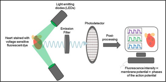
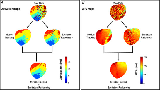
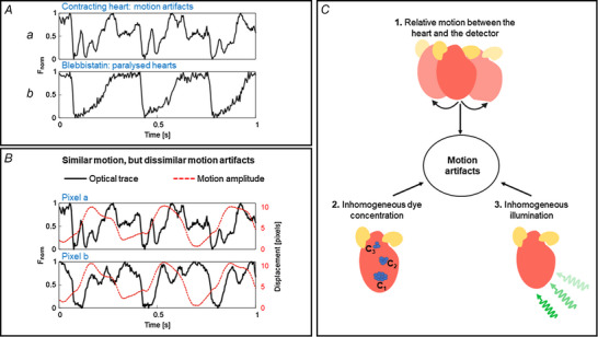
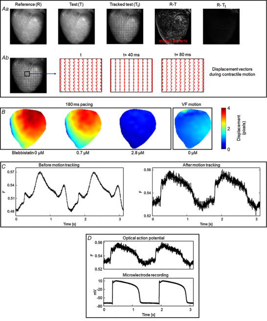
Optical mapping is a widely used tool to record and visualize the electrophysiological properties in a variety of myocardial preparations such as Langendorff-perfused isolated hearts, coronary-perfused wedge preparations, and cell culture monolayers. Motion artifact originating from the mechanical contraction of the myocardium creates a significant challenge to performing optical mapping of contracting hearts. Hence, to minimize the motion artifact, cardiac optical mapping studies are mostly performed on non-contracting hearts, where the mechanical contraction is removed using pharmacological excitation-contraction uncouplers. However, such experimental preparations eliminate the possibility of electromechanical interaction, and effects such as mechano-electric feedback cannot be studied. Recent developments in computer vision algorithms and ratiometric techniques have opened the possibility of performing optical mapping studies on isolated contracting hearts. In this review, we discuss the existing techniques and challenges of optical mapping of contracting hearts.
光学标测是一种广泛应用的工具,可用于记录和可视化各种心肌标本中的电生理特性,如 Langendorff 灌流分离心脏、冠状灌注楔形标本和细胞培养单层。源自心肌机械收缩的运动伪影给收缩心脏的光学标测带来了重大挑战。因此,为了最大程度地减少运动伪影,心脏光学标测研究主要在非收缩心脏上进行,通过使用药理学的兴奋-收缩偶联解偶联剂去除机械收缩。然而,这种实验制备消除了机电相互作用的可能性,并且无法研究机械电反馈等效应。计算机视觉算法和比率技术的最新发展为在分离的收缩心脏上进行光学标测研究开辟了可能性。在这篇综述中,我们讨论了收缩心脏光学标测的现有技术和挑战。