Department of Cardiovascular and Metabolic Sciences, Lerner Research Institute, Cleveland Clinic, Cleveland, OH.
Department of Biochemistry, Case Western Reserve University, Cleveland, OH.
Blood. 2023 May 25;141(21):2629-2641. doi: 10.1182/blood.2022018333.
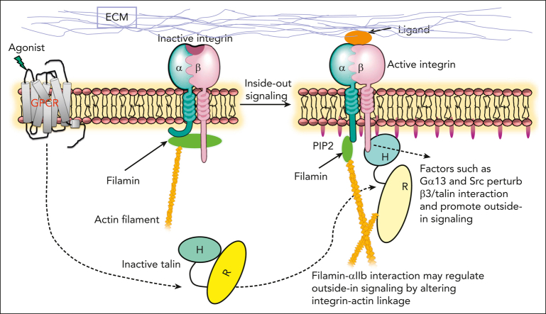
The communication of talin-activated integrin αIIbβ3 with the cytoskeleton (integrin outside-in signaling) is essential for platelet aggregation, wound healing, and hemostasis. Filamin, a large actin crosslinker and integrin binding partner critical for cell spreading and migration, is implicated as a key regulator of integrin outside-in signaling. However, the current dogma is that filamin, which stabilizes inactive αIIbβ3, is displaced from αIIbβ3 by talin to promote the integrin activation (inside-out signaling), and how filamin further functions remains unresolved. Here, we show that while associating with the inactive αIIbβ3, filamin also associates with the talin-bound active αIIbβ3 to mediate platelet spreading. Fluorescence resonance energy transfer-based analysis reveals that while associating with both αIIb and β3 cytoplasmic tails (CTs) to maintain the inactive αIIbβ3, filamin is spatiotemporally rearranged to associate with αIIb CT alone on activated αIIbβ3. Consistently, confocal cell imaging indicates that integrin α CT-linked filamin gradually delocalizes from the β CT-linked focal adhesion marker-vinculin likely because of the separation of integrin α/β CTs occurring during integrin activation. High-resolution crystal and nuclear magnetic resonance structure determinations unravel that the activated integrin αIIb CT binds to filamin via a striking α-helix→β-strand transition with a strengthened affinity that is dependent on the integrin-activating membrane environment containing enriched phosphatidylinositol 4,5-bisphosphate. These data suggest a novel integrin αIIb CT-filamin-actin linkage that promotes integrin outside-in signaling. Consistently, disruption of such linkage impairs the activation state of αIIbβ3, phosphorylation of focal adhesion kinase/proto-oncogene tyrosine kinase Src, and cell migration. Together, our findings advance the fundamental understanding of integrin outside-in signaling with broad implications in blood physiology and pathology.
衔接蛋白 talin 激活的整合素 αIIbβ3 与细胞骨架的相互作用(整合素外-内信号转导)对于血小板聚集、伤口愈合和止血至关重要。细丝蛋白(一种对细胞铺展和迁移至关重要的大肌动蛋白交联剂和整合素结合伴侣)被认为是整合素外-内信号转导的关键调节因子。然而,目前的主流观点认为,细丝蛋白稳定无活性的 αIIbβ3,通过衔接蛋白 talin 从 αIIbβ3 上置换下来,从而促进整合素的激活(内-外信号转导),但细丝蛋白如何进一步发挥作用仍未解决。在这里,我们表明,虽然与无活性的 αIIbβ3 结合,但细丝蛋白也与衔接蛋白结合的活性 αIIbβ3 结合,介导血小板铺展。荧光共振能量转移分析显示,尽管与 αIIb 和β3 的胞质尾部(CT)结合以维持无活性的 αIIbβ3,但细丝蛋白在空间和时间上被重新排列,仅与激活的 αIIbβ3 上的 αIIb CT 结合。一致地,共聚焦细胞成像表明,整合素α CT 连接的细丝蛋白逐渐从β CT 连接的粘着斑标志物 vinculin 上解聚,可能是由于整合素激活过程中整合素α/β CT 的分离。高分辨率晶体和核磁共振结构测定揭示,激活的整合素αIIb CT 通过一个引人注目的α-螺旋→β-链转变与细丝蛋白结合,亲和力增强,这取决于含有丰富的磷酸肌醇 4,5-二磷酸的整合素激活膜环境。这些数据表明了一种新的整合素αIIb CT-细丝蛋白-肌动蛋白连接,促进整合素外-内信号转导。一致地,破坏这种连接会损害 αIIbβ3 的激活状态、粘着斑激酶/原癌基因酪氨酸激酶Src 的磷酸化以及细胞迁移。总之,我们的发现推进了对整合素外-内信号转导的基本理解,对血液生理学和病理学具有广泛的意义。