Cetnar Ashley J, Degnan Michael, Pichler Joseph, Jain Sagarika, Morelli Samantha, Thomas Evan, Elder J Bradley, Scharschmidt Thomas J, Palmer Joshua D, Blakaj Dukagjin M
The Ohio State University, James Cancer Hospital and Solove Research Institute, Department of Radiation Oncology, 460 W. 10th Ave., Columbus, OH 43210, USA.
Phys Imaging Radiat Oncol. 2023 Feb 7;25:100422. doi: 10.1016/j.phro.2023.100422. eCollection 2023 Jan.
Mitigation of intrafraction motion (IM) is valuable in stereotactic radiotherapy (SRT) radiotherapy where submillimeter accuracy is desired. The purpose of this study was to investigate the application of triggered kilovoltage (kV) imaging for spine SRT patients with hardware by correlating kV imaging with patient motion and summarizing implications of tolerance for IM based on calculated dose.
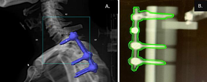
Ten plans (33 fractions) were studied, correlating kV imaging during treatment with pre- and post-treatment cone beam computed tomography (CBCT). Images were taken at 20-degree gantry angle intervals during the arc-based treatment. The contour of the hardware with a 1 mm expansion was displayed at the treatment console to manually pause treatment delivery if the hardware was visually detected outside the contour. The treatment CBCTs were compared using retrospective image registration to assess the validity of contour-based method for pausing treatment. Finally, plans were generated to estimate dose volume objective differences in case of 1 mm deviation.
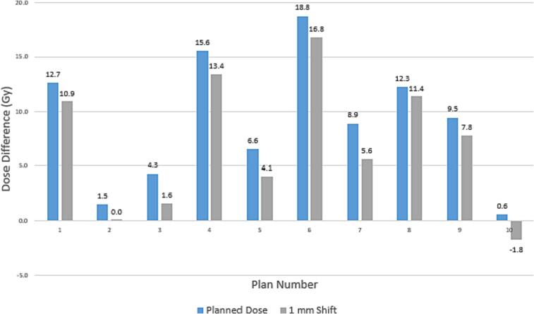
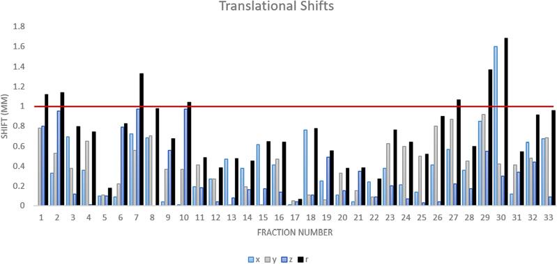
When kV imaging during treatment was used with the 1 mm contour, 100 % of the post-treatment CBCTs reported consistent results. One patient in the cohort exhibited motion greater than 1 mm during treatment which allowed intervention and re-setup during treatment. The average translational motion was 0.35 mm. Treatment plan comparison at 1 mm deviation showed little differences in calculated dose for the target and cord.
Utilizing kV imaging during treatment is an effective method of assessing IM for SRT spine patients with hardware without increasing treatment time.
在需要亚毫米精度的立体定向放射治疗(SRT)中,减轻分次内运动(IM)具有重要价值。本研究的目的是通过将千伏(kV)成像与患者运动相关联,并基于计算剂量总结IM耐受性的影响,来研究触发式kV成像在有植入物的脊柱SRT患者中的应用。
研究了10个计划(33次分次),将治疗期间的kV成像与治疗前和治疗后的锥形束计算机断层扫描(CBCT)相关联。在基于弧形的治疗过程中,以20度的机架角度间隔进行图像采集。如果在轮廓外肉眼检测到植入物,则在治疗控制台显示带有1毫米扩展的植入物轮廓,以手动暂停治疗输送。使用回顾性图像配准比较治疗CBCT,以评估基于轮廓的治疗暂停方法的有效性。最后,生成计划以估计在1毫米偏差情况下的剂量体积目标差异。
当在治疗期间使用带有1毫米轮廓的kV成像时,100%的治疗后CBCT报告结果一致。队列中的一名患者在治疗期间表现出大于1毫米的运动,这使得在治疗期间能够进行干预和重新设置。平均平移运动为0.35毫米。在1毫米偏差下的治疗计划比较显示,靶区和脊髓的计算剂量差异很小。
在治疗期间利用kV成像对于评估有植入物的脊柱SRT患者的IM是一种有效的方法,且不会增加治疗时间。