• 文献检索
  • 文档翻译
  • 深度研究
  • 学术资讯
  • Suppr Zotero 插件Zotero 插件
  • 邀请有礼
  • 套餐&价格
  • 历史记录
应用&插件
Suppr Zotero 插件Zotero 插件浏览器插件Mac 客户端Windows 客户端微信小程序
定价
高级版会员购买积分包购买API积分包
服务
文献检索文档翻译深度研究API 文档MCP 服务
关于我们
关于 Suppr公司介绍联系我们用户协议隐私条款
关注我们

Suppr 超能文献

核心技术专利:CN118964589B侵权必究
粤ICP备2023148730 号-1Suppr @ 2026

文献检索

告别复杂PubMed语法,用中文像聊天一样搜索,搜遍4000万医学文献。AI智能推荐,让科研检索更轻松。

立即免费搜索

文件翻译

保留排版,准确专业,支持PDF/Word/PPT等文件格式,支持 12+语言互译。

免费翻译文档

深度研究

AI帮你快速写综述,25分钟生成高质量综述,智能提取关键信息,辅助科研写作。

立即免费体验

所选MRI衍生的影像组学在鉴别中线胶质瘤和H3F3AK27M突变患者的无进展生存期和总生存期方面的诊断性能

Diagnostic Performance of Selected MRI-Derived Radiomics Able to Discriminate Progression-Free and Overall Survival in Patients with Midline Glioma and the H3F3AK27M Mutation.

作者信息

Chilaca-Rosas Maria-Fatima, Garcia-Lezama Melissa, Moreno-Jimenez Sergio, Roldan-Valadez Ernesto

机构信息

Radiotherapy Department, Hospital de Oncología, Centro Medico Nacional Siglo XXI, Instituto Mexicano del Seguro Social, Mexico City 06720, Mexico.

Directorate of Research, Hospital General de Mexico "Dr Eduardo Liceaga", Mexico City 06720, Mexico.

出版信息

Diagnostics (Basel). 2023 Feb 23;13(5):849. doi: 10.3390/diagnostics13050849.

DOI:10.3390/diagnostics13050849
PMID:36899993
原文链接:https://pmc.ncbi.nlm.nih.gov/articles/PMC10001394/
Abstract

BACKGROUND

Radiomics refers to a recent area of knowledge that studies features extracted from different imaging techniques and subsequently transformed into high-dimensional data that can be associated with biological events. Diffuse midline gliomas (DMG) are one of the most devastating types of cancer, with a median survival of approximately 11 months after diagnosis and 4-5 months after radiological and clinical progression.

METHODS

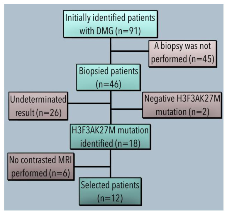
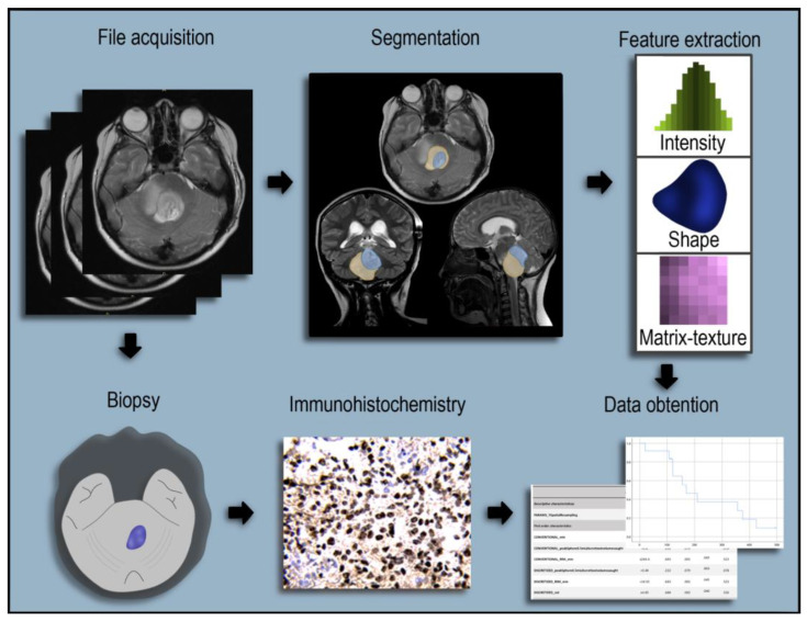
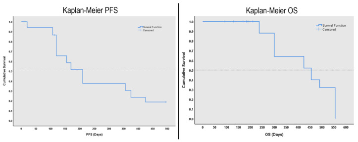
A retrospective study. From a database of 91 patients with DMG, only 12 had the H3.3K27M mutation and brain MRI DICOM files available. Radiomic features were extracted from MRI T1 and T2 sequences using LIFEx software. Statistical analysis included normal distribution tests and the Mann-Whitney U test, ROC analysis, and calculation of cut-off values.

RESULTS

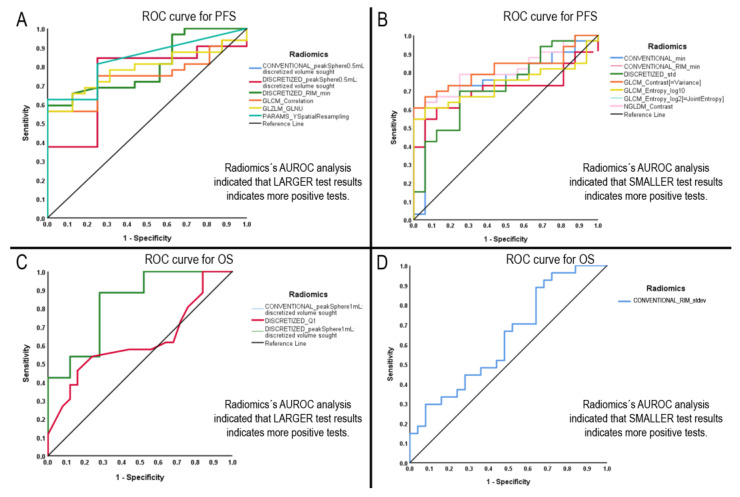
A total of 5760 radiomic values were included in the analyses. AUROC demonstrated 13 radiomics with statistical significance for progression-free survival (PFS) and overall survival (OS). Diagnostic performance tests showed nine radiomics with specificity for PFS above 90% and one with a sensitivity of 97.2%. For OS, 3 out of 4 radiomics demonstrated between 80 and 90% sensitivity.

CONCLUSIONS

Several radiomic features demonstrated statistical significance and have the potential to further aid DMG diagnostic assessment non-invasively. The most significant radiomics were first- and second-order features with GLCM texture profile, GLZLM_GLNU, and NGLDM_Contrast.

摘要

背景

放射组学是一个新兴的知识领域,它研究从不同成像技术中提取的特征,并随后将其转化为可与生物学事件相关联的高维数据。弥漫性中线胶质瘤(DMG)是最具毁灭性的癌症类型之一,诊断后的中位生存期约为11个月,放射学和临床进展后的中位生存期为4 - 5个月。

方法

一项回顾性研究。在一个包含91例DMG患者的数据库中,只有12例具有H3.3K27M突变且有脑部MRI DICOM文件。使用LIFEx软件从MRI T1和T2序列中提取放射组学特征。统计分析包括正态分布检验、曼 - 惠特尼U检验、ROC分析以及临界值计算。

结果

分析共纳入5760个放射组学值。AUROC显示有13个放射组学特征对无进展生存期(PFS)和总生存期(OS)具有统计学意义。诊断性能测试表明,9个放射组学特征对PFS的特异性高于90%,1个特征的敏感性为97.2%。对于OS,4个放射组学特征中有3个的敏感性在80%至90%之间。

结论

几个放射组学特征具有统计学意义,并且有可能进一步无创地辅助DMG的诊断评估。最显著的放射组学特征是具有GLCM纹理轮廓、GLZLM_GLNU和NGLDM_对比度的一阶和二阶特征。

https://cdn.ncbi.nlm.nih.gov/pmc/blobs/9f21/10001394/15a858b9b56d/diagnostics-13-00849-g005.jpg
https://cdn.ncbi.nlm.nih.gov/pmc/blobs/9f21/10001394/1eabf0b32946/diagnostics-13-00849-g001.jpg
https://cdn.ncbi.nlm.nih.gov/pmc/blobs/9f21/10001394/f14dd1f59c51/diagnostics-13-00849-g002.jpg
https://cdn.ncbi.nlm.nih.gov/pmc/blobs/9f21/10001394/9dbcff030628/diagnostics-13-00849-g003.jpg
https://cdn.ncbi.nlm.nih.gov/pmc/blobs/9f21/10001394/16726a608873/diagnostics-13-00849-g004.jpg
https://cdn.ncbi.nlm.nih.gov/pmc/blobs/9f21/10001394/15a858b9b56d/diagnostics-13-00849-g005.jpg

相似文献

1
Diagnostic Performance of Selected MRI-Derived Radiomics Able to Discriminate Progression-Free and Overall Survival in Patients with Midline Glioma and the H3F3AK27M Mutation.所选MRI衍生的影像组学在鉴别中线胶质瘤和H3F3AK27M突变患者的无进展生存期和总生存期方面的诊断性能
Diagnostics (Basel). 2023 Feb 23;13(5):849. doi: 10.3390/diagnostics13050849.
2
Identification of Radiomic Signatures in Brain MRI Sequences T1 and T2 That Differentiate Tumor Regions of Midline Gliomas with H3.3K27M Mutation.在T1和T2脑磁共振成像序列中鉴别区分具有H3.3K27M突变的中线胶质瘤肿瘤区域的影像组学特征
Diagnostics (Basel). 2023 Aug 14;13(16):2669. doi: 10.3390/diagnostics13162669.
3
Radiomic Features Based on MRI Predict Progression-Free Survival in Pediatric Diffuse Midline Glioma/Diffuse Intrinsic Pontine Glioma.基于MRI的影像组学特征预测儿童弥漫性中线胶质瘤/弥漫性脑桥内在型胶质瘤的无进展生存期
Can Assoc Radiol J. 2023 Feb;74(1):119-126. doi: 10.1177/08465371221109921. Epub 2022 Jun 29.
4
Exploring MRI Characteristics of Brain Diffuse Midline Gliomas With the H3 K27M Mutation Using Radiomics.利用放射组学探索具有H3 K27M突变的脑弥漫性中线胶质瘤的MRI特征
Front Oncol. 2021 May 24;11:646267. doi: 10.3389/fonc.2021.646267. eCollection 2021.
5
Development and validation of a machine learning algorithm for predicting diffuse midline glioma, H3 K27-altered, H3 K27 wild-type high-grade glioma, and primary CNS lymphoma of the brain midline in adults.开发和验证用于预测成人脑中线弥漫性中线胶质瘤、H3 K27 突变型、H3 K27 野生型高级别胶质瘤和原发性中枢神经系统淋巴瘤的机器学习算法。
J Neurosurg. 2022 Dec 23;139(2):393-401. doi: 10.3171/2022.11.JNS221544. Print 2023 Aug 1.
6
DCE-MRI Radiomics Analysis in Differentiating Luminal A and Luminal B Breast Cancer Molecular Subtypes.DCE-MRI 放射组学分析在鉴别 Luminal A 和 Luminal B 型乳腺癌分子亚型中的应用。
Acad Radiol. 2023 Jan;30(1):22-29. doi: 10.1016/j.acra.2022.04.004. Epub 2022 May 17.
7
Combination of clinical, radiomic, and "delta" radiomic features in survival prediction of metastatic gastroesophageal adenocarcinoma.临床、影像组学及“增量”影像组学特征在转移性胃食管腺癌生存预测中的联合应用
Front Oncol. 2023 Aug 14;13:892393. doi: 10.3389/fonc.2023.892393. eCollection 2023.
8
Predicting outcome in childhood diffuse midline gliomas using magnetic resonance imaging based texture analysis.使用基于磁共振成像的纹理分析预测儿童弥漫性中线胶质瘤的预后
J Neuroradiol. 2021 Jun;48(4):243-247. doi: 10.1016/j.neurad.2020.02.005. Epub 2020 Mar 14.
9
Physics-Informed Discretization for Reproducible and Robust Radiomic Feature Extraction Using Quantitative MRI.基于物理信息的离散化方法,用于使用定量 MRI 进行可重复且稳健的放射组学特征提取。
Invest Radiol. 2024 May 1;59(5):359-371. doi: 10.1097/RLI.0000000000001026. Epub 2023 Oct 9.
10
Dynamic susceptibility contrast-MRI parameters, ADC values, and the T2-FLAIR mismatch sign are useful to differentiate between H3-mutant and H3-wild-type high-grade midline glioma.动态磁敏感对比 MRI 参数、ADC 值和 T2-FLAIR 不匹配征象有助于鉴别 H3 突变型和 H3 野生型高级中线胶质瘤。
Eur Radiol. 2022 Jun;32(6):3672-3682. doi: 10.1007/s00330-021-08476-7. Epub 2022 Jan 13.

引用本文的文献

1
Systematic review and epistemic meta-analysis to advance binomial AI-radiomics integration for predicting high-grade glioma progression and enhancing patient management.系统评价与认知元分析,以推进二项式人工智能-放射组学整合,用于预测高级别胶质瘤进展并改善患者管理。
Sci Rep. 2025 May 8;15(1):16113. doi: 10.1038/s41598-025-98058-0.
2
MRI radiomics based on machine learning in high-grade gliomas as a promising tool for prediction of CD44 expression and overall survival.基于机器学习的MRI影像组学在高级别胶质瘤中作为预测CD44表达和总生存期的一种有前景的工具。
Sci Rep. 2025 Mar 3;15(1):7433. doi: 10.1038/s41598-025-90128-7.
3
Radiomics and artificial intelligence applications in pediatric brain tumors.

本文引用的文献

1
A meta-analysis of based radiomics for predicting lymph node metastasis in patients with biliary tract cancers.基于放射组学预测胆道癌患者淋巴结转移的荟萃分析。
Front Surg. 2023 Jan 6;9:1045295. doi: 10.3389/fsurg.2022.1045295. eCollection 2022.
2
Current status and future perspectives of radiomics in hepatocellular carcinoma.肝癌放射组学的现状与展望。
World J Gastroenterol. 2023 Jan 7;29(1):43-60. doi: 10.3748/wjg.v29.i1.43.
3
Machine learning with textural analysis of longitudinal multiparametric MRI and molecular subtypes accurately predicts pathologic complete response in patients with invasive breast cancer.
放射组学和人工智能在小儿脑肿瘤中的应用。
World J Pediatr. 2024 Aug;20(8):747-763. doi: 10.1007/s12519-024-00823-0. Epub 2024 Jun 27.
4
The value of MRI-based radiomics for evaluating early parotid gland injury in primary Sjögren's syndrome.基于MRI的影像组学在评估原发性干燥综合征早期腮腺损伤中的价值。
Clin Rheumatol. 2024 May;43(5):1675-1682. doi: 10.1007/s10067-024-06935-2. Epub 2024 Mar 27.
5
Identification of Radiomic Signatures in Brain MRI Sequences T1 and T2 That Differentiate Tumor Regions of Midline Gliomas with H3.3K27M Mutation.在T1和T2脑磁共振成像序列中鉴别区分具有H3.3K27M突变的中线胶质瘤肿瘤区域的影像组学特征
Diagnostics (Basel). 2023 Aug 14;13(16):2669. doi: 10.3390/diagnostics13162669.
基于纹理分析的纵向多参数 MRI 与分子亚型的机器学习可准确预测浸润性乳腺癌患者的病理完全缓解。
PLoS One. 2023 Jan 17;18(1):e0280320. doi: 10.1371/journal.pone.0280320. eCollection 2023.
4
Paediatric-type diffuse high-grade gliomas in the 5th CNS WHO Classification.第五版 CNS WHO 分类中的小儿型弥漫性高级别神经胶质瘤。
Pathologica. 2022 Dec;114(6):422-435. doi: 10.32074/1591-951X-830.
5
Interdisciplinary care of children with diffuse midline glioma.弥漫性中线胶质瘤患儿的多学科护理。
Neoplasia. 2023 Jan;35:100851. doi: 10.1016/j.neo.2022.100851. Epub 2022 Nov 18.
6
Predicting overall survival in diffuse glioma from the presurgical connectome.从术前连接组预测弥漫性神经胶质瘤的总生存期。
Sci Rep. 2022 Nov 5;12(1):18783. doi: 10.1038/s41598-022-22387-7.
7
Radiomics in neuro-oncological clinical trials.神经肿瘤学临床试验中的放射组学。
Lancet Digit Health. 2022 Nov;4(11):e841-e849. doi: 10.1016/S2589-7500(22)00144-3. Epub 2022 Sep 28.
8
Pediatric Diffuse Midline Gliomas: An Unfinished Puzzle.小儿弥漫性中线胶质瘤:一个未完成的谜题。
Diagnostics (Basel). 2022 Aug 25;12(9):2064. doi: 10.3390/diagnostics12092064.
9
Ultrasound radiomics in personalized breast management: Current status and future prospects.个性化乳腺管理中的超声影像组学:现状与未来展望
Front Oncol. 2022 Aug 17;12:963612. doi: 10.3389/fonc.2022.963612. eCollection 2022.
10
Discriminators of pseudoprogression and true progression in high-grade gliomas: A systematic review and meta-analysis.高级别胶质瘤中假性进展和真性进展的鉴别因素:系统评价和荟萃分析。
Sci Rep. 2022 Aug 2;12(1):13258. doi: 10.1038/s41598-022-16726-x.