Ibad Hamza Ahmed, Kwee Robert M, Ghotbi Elena, Roemer Frank W, Guermazi Ali, Demehri Shadpour
The Russell H. Morgan Department of Radiology and Radiological Science, Johns Hopkins University School of Medicine, Baltimore, MD, USA.
Department of Radiology, Zuyderland Medical Center, Heerlen/Sittard/Geleen, the Netherlands.
Osteoarthr Cartil Open. 2023 Feb 23;5(2):100348. doi: 10.1016/j.ocarto.2023.100348. eCollection 2023 Jun.
To determine the association between Intra-articular mineralization (IAM) and knee osteoarthritis (OA) outcomes stratified according to participants' age.
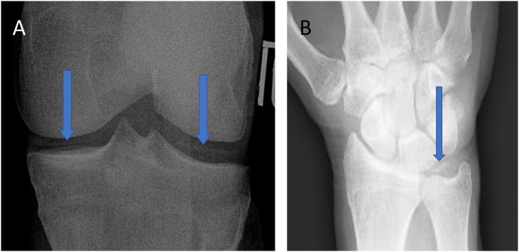
Participants from the Osteoarthritis Initiative (OAI) with baseline radiographic OA (i.e., Kellgren-Lawrence grade ≥2 with Osteoarthritis Research Society International (OARSI) atlas joint space narrowing (JSN)) in either knee were identified. Both knees and dominant hand baseline radiographs were evaluated for the presence of IAM. Whole-grade OARSI-JSN radiographic progression and increased Western Ontario and McMaster universities osteoarthritis index scores of the knees with baseline radiographic OA (assessed annually) were defined as radiographic and symptomatic progression, respectively. Cox proportional-hazards and longitudinal multilevel regression models investigated radiographic and symptomatic progression, respectively.
2010 participants with baseline radiographic OA in either one or both knees (N = 2976) were identified. 178 participants had baseline IAM (hand radiographs = 46, knee radiographs = 166, both = 34). An adjusted logistic regression model suggests an association between age and IAM (Odds Ratio: 1.06, 95% Confidence Interval (CI): 1.04-1.08). Presence of any IAM was not associated with whole-grade OARSI-JSN (Hazard Ratio (HR): 1.00, 95% CI: 0.73-1.37) or symptomatic progression (Estimated difference: 1.24, p-value: 0.13) in all participants. Using stratification analysis, in younger participants <60 years old, presence of any IAM was associated with radiographic progression (HR: 1.90, 95% CI: 1.01-3.60).
Although the presence of any radiographic IAM increases with higher age and does not predict knee OA outcomes across the entire sample of OAI participants, it is associated with knee OA radiographic progression in participants aged <60.
确定关节内矿化(IAM)与根据参与者年龄分层的膝关节骨关节炎(OA)结局之间的关联。
确定骨关节炎倡议组织(OAI)中任一侧膝关节有基线放射学OA(即,根据国际骨关节炎研究学会(OARSI)图谱,凯尔格伦-劳伦斯分级≥2且关节间隙变窄(JSN))的参与者。评估双膝和优势手的基线X线片是否存在IAM。全等级OARSI-JSN放射学进展以及基线放射学OA的膝关节(每年评估)的西安大略和麦克马斯特大学骨关节炎指数评分增加分别定义为放射学进展和症状性进展。Cox比例风险模型和纵向多水平回归模型分别研究放射学进展和症状性进展。
确定了2010名任一侧或双侧膝关节有基线放射学OA的参与者(N = 2976)。178名参与者有基线IAM(手部X线片 = 46,膝关节X线片 = 166,两者皆有 = 34)。校正后的逻辑回归模型表明年龄与IAM之间存在关联(优势比:1.06,95%置信区间(CI):1.04 - 1.08)。在所有参与者中,任何IAM的存在与全等级OARSI-JSN(风险比(HR):1.00,95%CI:0.73 - 1.37)或症状性进展(估计差异:1.24,p值:0.13)均无关联。使用分层分析,在年龄小于60岁的较年轻参与者中,任何IAM的存在与放射学进展相关(HR:1.90,95%CI:1.01 - 3.60)。
尽管任何放射学IAM的存在随年龄增长而增加,并且不能预测OAI参与者整个样本中的膝关节OA结局,但它与年龄小于60岁的参与者的膝关节OA放射学进展相关。