Castor Delivette, Saidu Rakiya, Boa Rosalind, Mbatani Nomonde, Mutsvangwa Tinashe E M, Moodley Jennifer, Denny Lynette, Kuhn Louise
Division of Infectious Diseases, Vagelos College of Physicians and Surgeons, Columbia University Irving Medical Center, New York, NY, United States.
Department of Epidemiology, Mailman School of Public Health, Columbia University Irving Medical Center, New York, NY, United States.
Front Health Serv. 2022 Sep 12;2:1000150. doi: 10.3389/frhs.2022.1000150. eCollection 2022.
We assessed the implementation context and image quality in preparation for a clinical study evaluating the effectiveness of automated visual assessment devices within cervical cancer screening of women living without and with HIV.
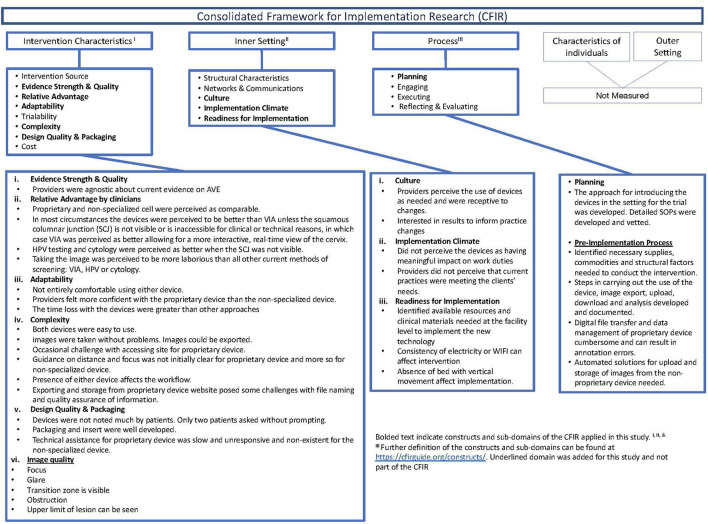
We developed a semi-structured questionnaire based on three Consolidated Framework for Implementation Research (CFIR) domains; intervention characteristics, inner setting, and process, in Cape Town, South Africa. Between December 1, 2020, and August 6, 2021, we evaluated two devices: MobileODT handheld colposcope; and a commercially-available cell phone (Samsung A21ST). Colposcopists visually inspected cervical images for technical adequacy. Descriptive analyses were tabulated for quantitative variables, and narrative responses were summarized in the text.
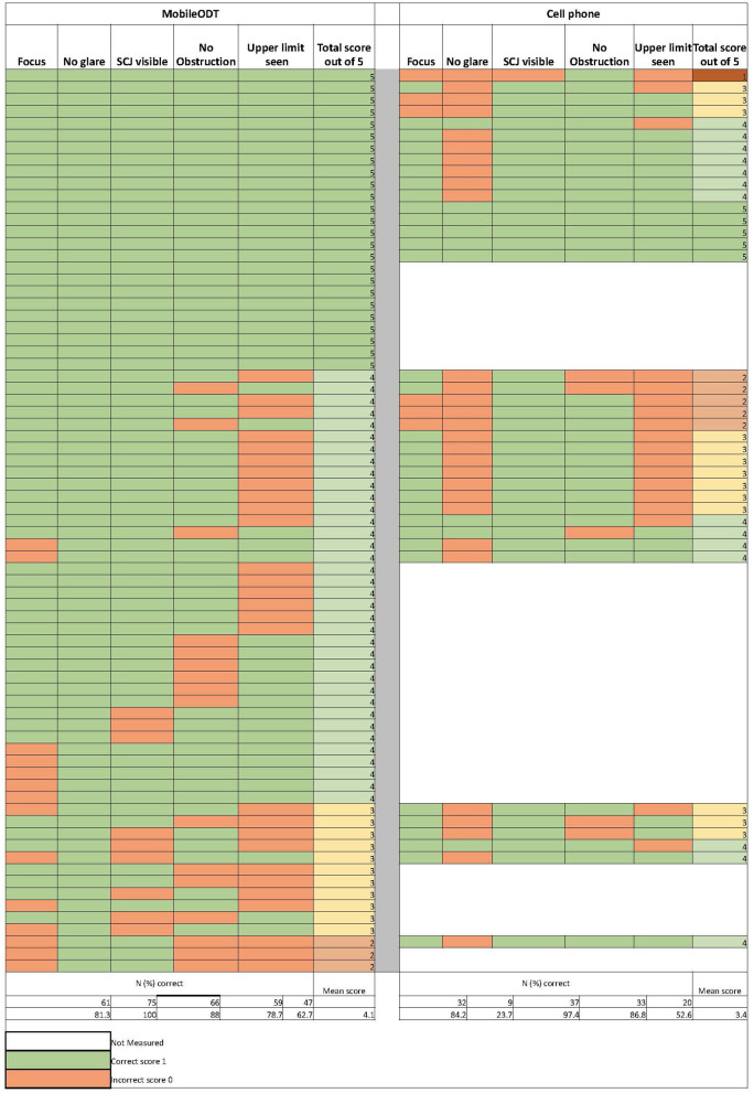
Two colposcopists described the devices as easy to operate, without data loss. The clinical workspace and gynecological workflow were modified to incorporate devices and manage images. Providers believed either device would likely perform better than cytology under most circumstances unless the squamocolumnar junction (SCJ) were not visible, in which case cytology was expected to be better. Image quality ( = 75) from the MobileODT device and cell phone was comparable in terms of achieving good focus (81% vs. 84%), obtaining visibility of the squamous columnar junction (88% vs. 97%), avoiding occlusion (79% vs. 87%), and detection of lesion and range of lesion includes the upper limit (63% vs. 53%) but differed in taking photographs free of glare (100% vs. 24%).
Novel application of the CFIR early in the conduct of the clinical study, including assessment of image quality, highlight real-world factors about intervention characteristics, inner clinical setting, and workflow process that may affect both the clinical study findings and ultimate pace of translating to clinical practice. The application and augmentation of the CFIR in this study context highlighted adaptations needed for the framework to better measure factors relevant to implementing digital interventions.
我们评估了实施背景和图像质量,为一项临床研究做准备,该研究旨在评估自动视觉评估设备在未感染和感染艾滋病毒的女性宫颈癌筛查中的有效性。
我们在南非开普敦,基于实施研究综合框架(CFIR)的三个领域(干预特征、内部环境和过程)编制了一份半结构化问卷。在2020年12月1日至2021年8月6日期间,我们评估了两种设备:MobileODT手持阴道镜;以及一款商用手机(三星A21ST)。阴道镜检查人员对宫颈图像进行视觉检查,以评估技术是否充分。对定量变量进行描述性分析并制成表格,文本中总结了叙述性回答。
两名阴道镜检查人员称这些设备易于操作,且无数据丢失情况。临床工作区和妇科工作流程进行了修改,以纳入设备并管理图像。提供者认为,在大多数情况下,除非鳞柱交界(SCJ)不可见,否则这两种设备可能比细胞学检查表现更好,在鳞柱交界不可见的情况下,预计细胞学检查效果会更好。MobileODT设备和手机的图像质量(n = 75)在实现良好对焦(81%对84%)、获得鳞柱交界的可见性(88%对97%)、避免遮挡(79%对87%)以及检测病变和病变范围包括上限(63%对53%)方面相当,但在拍摄无眩光照片方面有所不同(100%对24%)。
在临床研究早期对CFIR的新颖应用,包括图像质量评估,凸显了关于干预特征、内部临床环境和工作流程的现实因素,这些因素可能影响临床研究结果以及最终转化为临床实践的速度。CFIR在本研究背景下的应用和扩充凸显了该框架为更好地衡量与实施数字干预相关因素所需的调整。