Boo Jihyeon, Lee Jongwon, Kim Young-Hyun, Lee Chang-Hee, Ku Bonsu, Shin Injae
Department of Chemistry, Yonsei University, Seoul, Republic of Korea.
Disease Target Structure Research Center, Korea Research Institute of Bioscience and Biotechnology (KRIBB), Daejeon, Republic of Korea.
Front Chem. 2023 Mar 1;11:1133018. doi: 10.3389/fchem.2023.1133018. eCollection 2023.
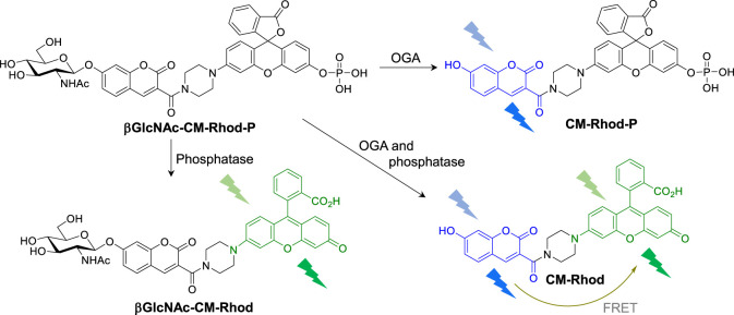
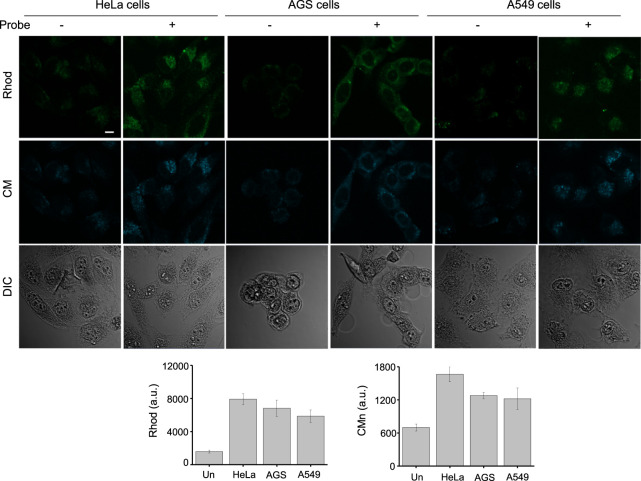
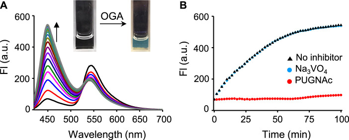
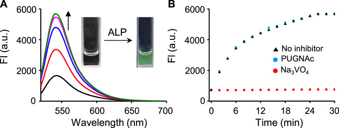
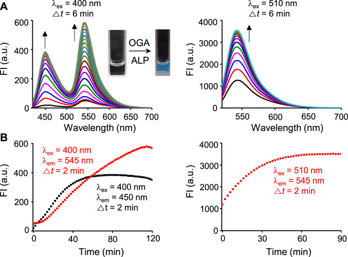
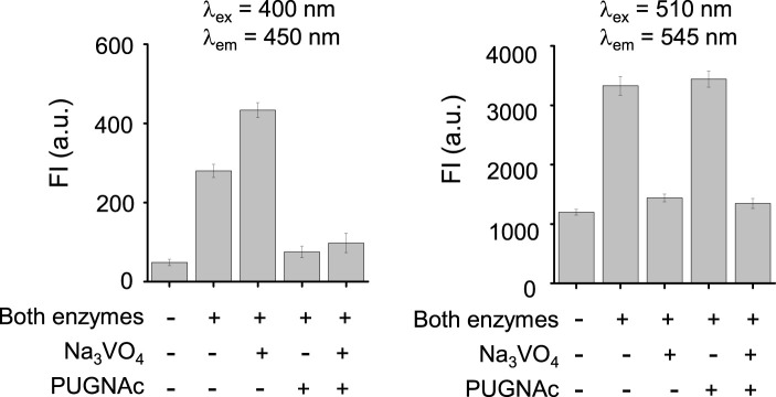
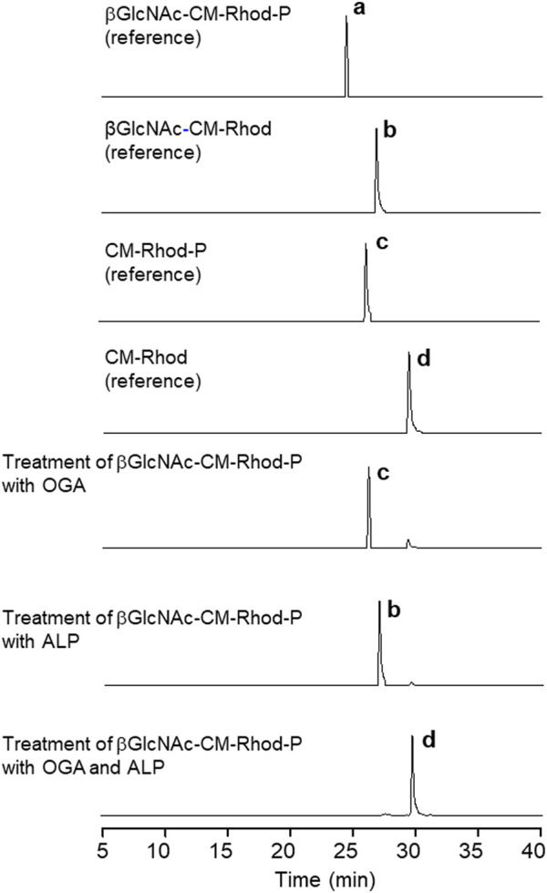
O-GlcNAc modification of proteins often has crosstalk with protein phosphorylation. These posttranslational modifications are highly dynamic events that modulate a wide range of cellular processes. Owing to the physiological and pathological significance of protein O-GlcNAcylation and phosphorylation, we designed the fluorescent probe, βGlcNAc-CM-Rhod-P, to differentially detect activities of O-GlcNAcase (OGA) and phosphatase, enzymes that are responsible for these modifications. βGlcNAc-CM-Rhod-P was comprised of a βGlcNAc-conjugated coumarin (βGlcNAc-CM) acting as an OGA substrate, a phosphorylated rhodol (Rhod-P) as a phosphatase substrate and a piperazine bridge. Because the emission wavelength maxima of CM and Rhod liberated from the probe are greatly different (100 nm), spectral interference is avoided. The results of this study revealed that treatment of βGlcNAc-CM-Rhod-P with OGA promotes formation of the GlcNAc-cleaved probe, CM-Rhod-P, and a consequent increase in the intensity of fluorescence associated with free CM. Also, it was found that exposure of the probe to phosphatase produces a dephosphorylated probe, βGlcNAc-CM-Rhod, which displays strong fluorescence arising from free Rhod. On the other hand, when incubated with both OGA and phosphatase, βGlcNAc-CM-Rhod-P was converted to CM-Rhod which lacked both βGlcNAc and phosphoryl groups, in conjunction with increases in the intensities of fluorescence arising from both free CM and Rhod. This probe was employed to detect activities of OGA and phosphatase in cell lysates and to fluorescently image both enzymes in cells. Collectively, the findings indicate that βGlcNAc-CM-Rhod-P can be utilized as a chemical tool to simultaneously determine activities of OGA and phosphatase.
蛋白质的O-连接N-乙酰葡糖胺(O-GlcNAc)修饰常常与蛋白质磷酸化存在相互作用。这些翻译后修饰是高度动态的事件,可调节广泛的细胞过程。鉴于蛋白质O-GlcNAc化和磷酸化在生理和病理方面的重要性,我们设计了荧光探针βGlcNAc-CM-Rhod-P,用于差异检测O-连接N-乙酰葡糖胺酶(OGA)和磷酸酶的活性,这两种酶分别负责这些修饰。βGlcNAc-CM-Rhod-P由作为OGA底物的βGlcNAc共轭香豆素(βGlcNAc-CM)、作为磷酸酶底物的磷酸化玫红酚(Rhod-P)以及哌嗪桥组成。由于从探针释放的CM和Rhod的最大发射波长差异很大(100nm),因此避免了光谱干扰。本研究结果表明,用OGA处理βGlcNAc-CM-Rhod-P会促进GlcNAc切割探针CM-Rhod-P的形成,进而导致与游离CM相关的荧光强度增加。此外,还发现将探针暴露于磷酸酶会产生去磷酸化探针βGlcNAc-CM-Rhod,其显示出由游离Rhod产生的强荧光。另一方面,当与OGA和磷酸酶一起孵育时,βGlcNAc-CM-Rhod-P会转化为既缺乏βGlcNAc又缺乏磷酸基团的CM-Rhod,同时游离CM和Rhod产生的荧光强度都会增加。该探针用于检测细胞裂解物中OGA和磷酸酶的活性,并对细胞中的这两种酶进行荧光成像。总的来说,这些发现表明βGlcNAc-CM-Rhod-P可作为一种化学工具,用于同时测定OGA和磷酸酶的活性。