Adey Brett N, Cooper-Knock Johnathan, Al Khleifat Ahmad, Fogh Isabella, van Damme Philip, Corcia Philippe, Couratier Philippe, Hardiman Orla, McLaughlin Russell, Gotkine Marc, Drory Vivian, Silani Vincenzo, Ticozzi Nicola, Veldink Jan H, van den Berg Leonard H, de Carvalho Mamede, Pinto Susana, Mora Pardina Jesus S, Povedano Panades Mónica, Andersen Peter M, Weber Markus, Başak Nazli A, Shaw Christopher E, Shaw Pamela J, Morrison Karen E, Landers John E, Glass Jonathan D, Vourc'h Patrick, Dobson Richard J B, Breen Gerome, Al-Chalabi Ammar, Jones Ashley R, Iacoangeli Alfredo
Social Genetic and Developmental Psychiatry Centre, Institute of Psychiatry, Psychology and Neuroscience, King's College London, London, United Kingdom.
Department of Biostatistics and Health Informatics, Institute of Psychiatry, Psychology and Neuroscience, King's College London, London, United Kingdom.
Front Cell Neurosci. 2023 Mar 2;17:1112405. doi: 10.3389/fncel.2023.1112405. eCollection 2023.
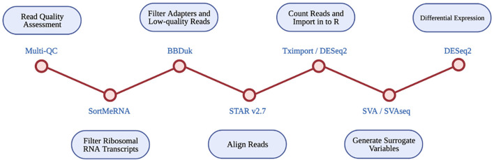
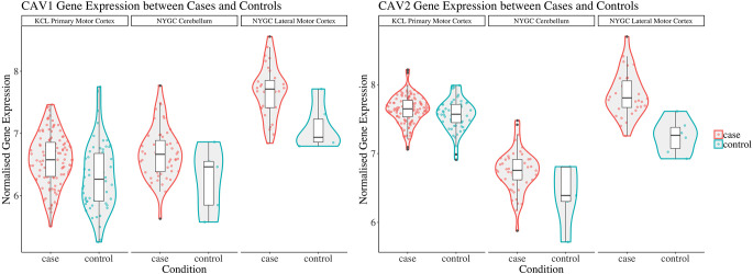
Caveolin-1 and Caveolin-2 (CAV1 and CAV2) are proteins associated with intercellular neurotrophic signalling. There is converging evidence that CAV1 and CAV2 (CAV1/2) genes have a role in amyotrophic lateral sclerosis (ALS). Disease-associated variants have been identified within CAV1/2 enhancers, which reduce gene expression and lead to disruption of membrane lipid rafts. Using large ALS whole-genome sequencing and post-mortem RNA sequencing datasets (5,987 and 365 tissue samples, respectively), and iPSC-derived motor neurons from 55 individuals, we investigated the role of CAV1/2 expression and enhancer variants in the ALS phenotype. We report a differential expression analysis between ALS cases and controls for CAV1 and CAV2 genes across various post-mortem brain tissues and three independent datasets. CAV1 and CAV2 expression was consistently higher in ALS patients compared to controls, with significant results across the primary motor cortex, lateral motor cortex, and cerebellum. We also identify increased survival among carriers of CAV1/2 enhancer mutations compared to non-carriers within Project MinE and slower progression as measured by the ALSFRS. Carriers showed a median increase in survival of 345 days. These results add to an increasing body of evidence linking CAV1 and CAV2 genes to ALS. We propose that carriers of CAV1/2 enhancer mutations may be conceptualised as an ALS subtype who present a less severe ALS phenotype with a longer survival duration and slower progression. Upregulation of CAV1/2 genes in ALS cases may indicate a causal pathway or a compensatory mechanism. Given prior research supporting the beneficial role of CAV1/2 expression in ALS patients, we consider a compensatory mechanism to better fit the available evidence, although further investigation into the biological pathways associated with CAV1/2 is needed to support this conclusion.
小窝蛋白-1和小窝蛋白-2(CAV1和CAV2)是与细胞间神经营养信号传导相关的蛋白质。越来越多的证据表明,CAV1和CAV2(CAV1/2)基因在肌萎缩侧索硬化症(ALS)中起作用。已在CAV1/2增强子内鉴定出与疾病相关的变体,这些变体降低基因表达并导致膜脂筏的破坏。利用大型ALS全基因组测序和死后RNA测序数据集(分别为5987个和365个组织样本),以及来自55名个体的诱导多能干细胞衍生的运动神经元,我们研究了CAV1/2表达和增强子变体在ALS表型中的作用。我们报告了在各种死后脑组织和三个独立数据集中,ALS病例与对照之间CAV1和CAV2基因的差异表达分析。与对照相比,ALS患者中CAV1和CAV2的表达始终更高,在初级运动皮层、外侧运动皮层和小脑中均有显著结果。我们还发现,与“最小化工程”项目中的非携带者相比,CAV1/2增强子突变携带者的生存期延长,并且根据肌萎缩侧索硬化功能评定量表(ALSFRS)测量,疾病进展较慢。携带者的生存期中位数增加了345天。这些结果进一步证明了CAV1和CAV2基因与ALS之间的联系。我们提出,CAV1/2增强子突变携带者可被视为一种ALS亚型,其表现出不太严重的ALS表型,生存期更长,进展更慢。ALS病例中CAV1/2基因的上调可能表明一种因果途径或一种补偿机制。鉴于先前的研究支持CAV1/2表达在ALS患者中的有益作用,我们认为补偿机制更符合现有证据,尽管需要进一步研究与CAV1/2相关的生物学途径来支持这一结论。