• 文献检索
  • 文档翻译
  • 深度研究
  • 学术资讯
  • Suppr Zotero 插件Zotero 插件
  • 邀请有礼
  • 套餐&价格
  • 历史记录
应用&插件
Suppr Zotero 插件Zotero 插件浏览器插件Mac 客户端Windows 客户端微信小程序
定价
高级版会员购买积分包购买API积分包
服务
文献检索文档翻译深度研究API 文档MCP 服务
关于我们
关于 Suppr公司介绍联系我们用户协议隐私条款
关注我们

Suppr 超能文献

核心技术专利:CN118964589B侵权必究
粤ICP备2023148730 号-1Suppr @ 2026

文献检索

告别复杂PubMed语法,用中文像聊天一样搜索,搜遍4000万医学文献。AI智能推荐,让科研检索更轻松。

立即免费搜索

文件翻译

保留排版,准确专业,支持PDF/Word/PPT等文件格式,支持 12+语言互译。

免费翻译文档

深度研究

AI帮你快速写综述,25分钟生成高质量综述,智能提取关键信息,辅助科研写作。

立即免费体验

扩张的血管周围间隙是多发性硬化症中动脉疾病的一个标志物。

Dilated Virchow-Robin Spaces are a Marker for Arterial Disease in Multiple Sclerosis.

作者信息

Ineichen Benjamin V, Cananau Carmen, Plattén Michael, Ouellette Russell, Moridi Thomas, Frauenknecht Katrin B M, Okar Serhat V, Kulcsar Zsolt, Kockum Ingrid, Piehl Fredrik, Reich Daniel S, Granberg Tobias

机构信息

Department of Clinical Neuroscience, Karolinska Institutet, Stockholm, Sweden.

Department of Neuroradiology, Karolinska University Hospital, Stockholm, Sweden.

出版信息

bioRxiv. 2023 Feb 27:2023.02.24.529871. doi: 10.1101/2023.02.24.529871.

DOI:10.1101/2023.02.24.529871
PMID:36945422
原文链接:https://pmc.ncbi.nlm.nih.gov/articles/PMC10028816/
Abstract

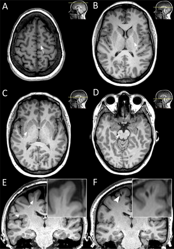
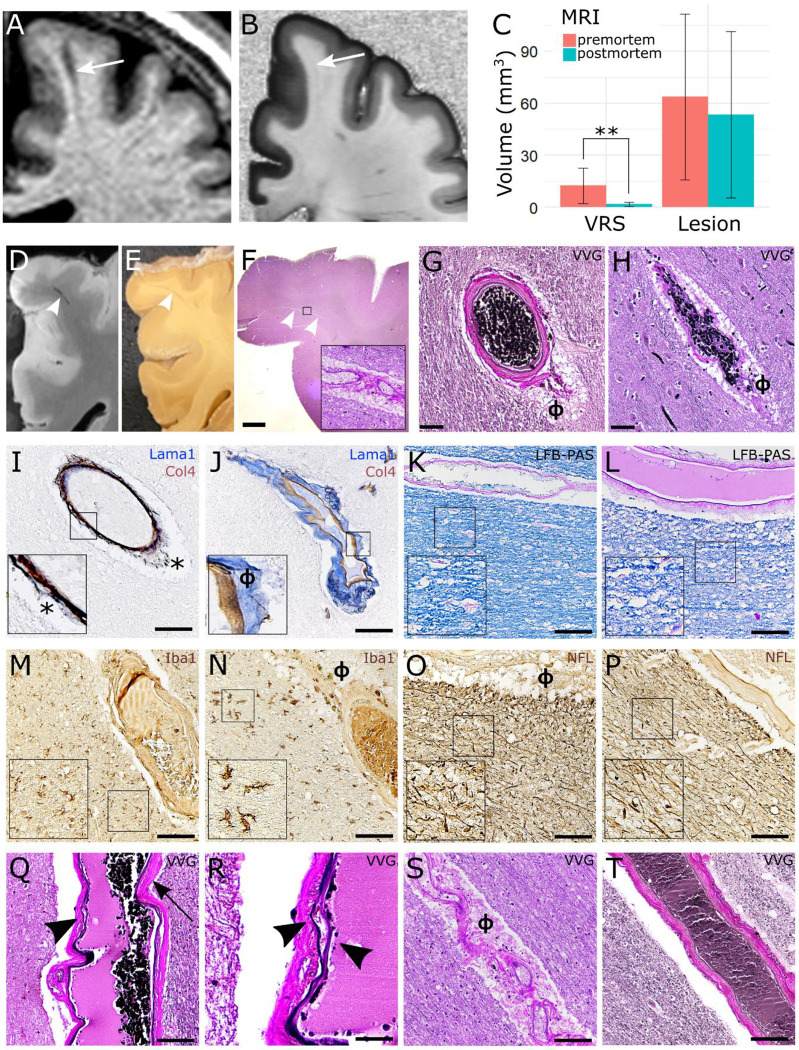
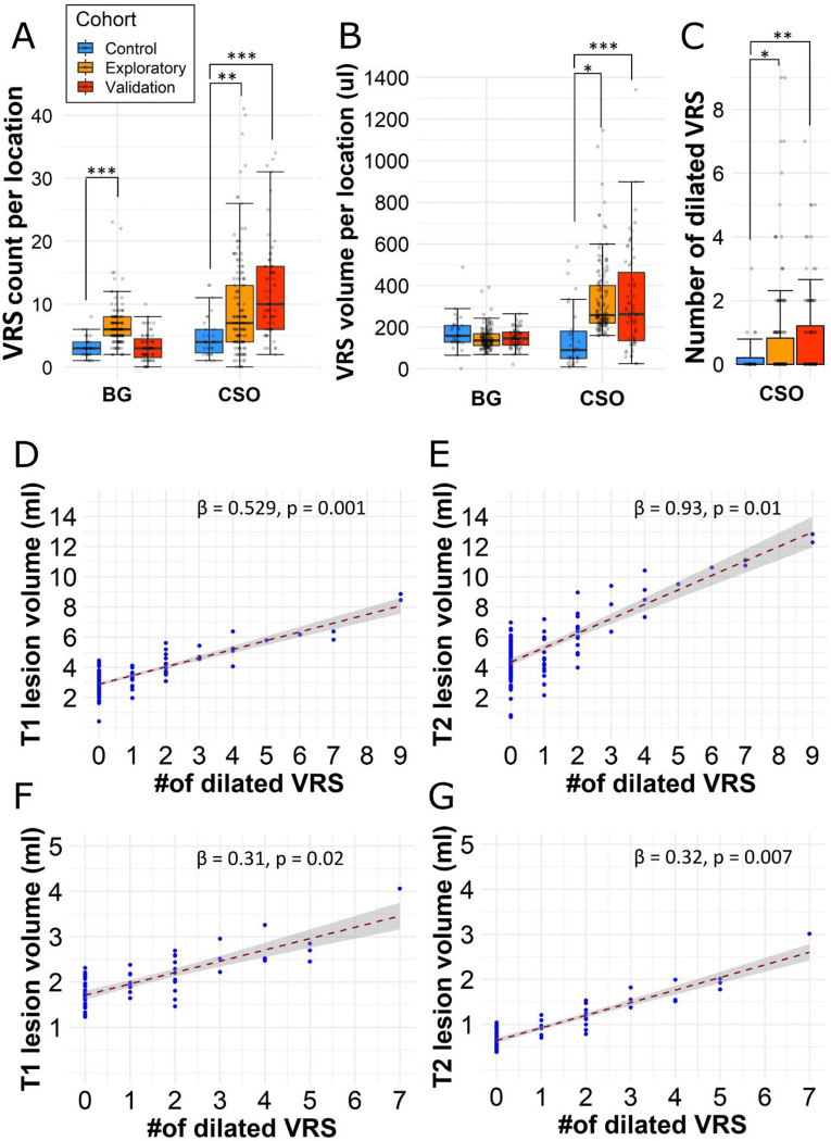
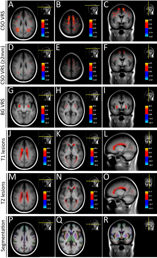
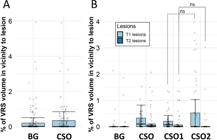
Virchow-Robin spaces (VRS) have been associated with neurodegeneration and neuroinflammation. However, it remains uncertain to what degree non-dilated or dilated VRS reflect specific features of neuroinflammatory pathology. Thus, we aimed at investigating the clinical relevance of VRS as imaging biomarker in multiple sclerosis (MS) and to correlate VRS to their histopathologic signature. In a cohort study comprising 205 MS patients (including a validation cohort) and 30 control subjects, we assessed the association of non-dilated and dilated VRS to clinical and magnetic resonance imaging (MRI) out-comes. Brain blocks from 6 MS patients and 3 non-MS controls were histopathologically processed to correlate VRS to their tissue substrate. The count of dilated centrum semiovale VRS was associated with increased T1 and T2 lesion volumes. There was no systematic spatial colocalization of dilated VRS with MS lesions. At tissue level, VRS mostly corresponded to arteries and were not associated with MS pathological hallmarks. Interestingly, dilated VRS in MS were associated with signs of small vessel disease. Contrary to prior beliefs, these observations suggest that VRS in MS do not associate with accumulation of immune cells. But instead, these findings indicate vascular pathology as a driver and/or consequence of neuroinflammatory pathology for this imaging feature.

摘要

血管周围间隙(VRS)与神经退行性变和神经炎症有关。然而,非扩张性或扩张性VRS在多大程度上反映神经炎症病理的特定特征仍不确定。因此,我们旨在研究VRS作为多发性硬化症(MS)成像生物标志物的临床相关性,并将VRS与其组织病理学特征相关联。在一项包括205名MS患者(包括一个验证队列)和30名对照受试者的队列研究中,我们评估了非扩张性和扩张性VRS与临床和磁共振成像(MRI)结果的关联。对6名MS患者和3名非MS对照的脑块进行组织病理学处理,以将VRS与其组织基质相关联。扩张的半卵圆中心VRS计数与T1和T2病变体积增加相关。扩张的VRS与MS病变没有系统性的空间共定位。在组织水平上,VRS大多与动脉相对应,与MS病理特征无关。有趣的是,MS中扩张的VRS与小血管疾病的体征相关。与先前的观点相反,这些观察结果表明,MS中的VRS与免疫细胞的积累无关。但相反,这些发现表明血管病理是这种成像特征的神经炎症病理的驱动因素和/或结果。

https://cdn.ncbi.nlm.nih.gov/pmc/blobs/5208/10028816/3f860ed789d1/nihpp-2023.02.24.529871v1-f0005.jpg
https://cdn.ncbi.nlm.nih.gov/pmc/blobs/5208/10028816/e9aa4c48f69e/nihpp-2023.02.24.529871v1-f0001.jpg
https://cdn.ncbi.nlm.nih.gov/pmc/blobs/5208/10028816/b2007f414724/nihpp-2023.02.24.529871v1-f0002.jpg
https://cdn.ncbi.nlm.nih.gov/pmc/blobs/5208/10028816/936ca29af581/nihpp-2023.02.24.529871v1-f0003.jpg
https://cdn.ncbi.nlm.nih.gov/pmc/blobs/5208/10028816/3fd36b6cbb49/nihpp-2023.02.24.529871v1-f0004.jpg
https://cdn.ncbi.nlm.nih.gov/pmc/blobs/5208/10028816/3f860ed789d1/nihpp-2023.02.24.529871v1-f0005.jpg

相似文献

1
Dilated Virchow-Robin Spaces are a Marker for Arterial Disease in Multiple Sclerosis.扩张的血管周围间隙是多发性硬化症中动脉疾病的一个标志物。
bioRxiv. 2023 Feb 27:2023.02.24.529871. doi: 10.1101/2023.02.24.529871.
2
Dilated Virchow-Robin spaces are a marker for arterial disease in multiple sclerosis.扩大的血管周围间隙是多发性硬化症中动脉疾病的一个标志物。
EBioMedicine. 2023 Jun;92:104631. doi: 10.1016/j.ebiom.2023.104631. Epub 2023 May 28.
3
Perivascular spaces in MS patients at 7 Tesla MRI: a marker of neurodegeneration?7特斯拉MRI下MS患者的血管周围间隙:神经退行性变的标志物?
Mult Scler. 2015 Feb;21(2):155-62. doi: 10.1177/1352458514540358. Epub 2014 Jul 10.
4
Dilated perivascular spaces can present incidental CSF-isointense foci within the ventral forebrain of dogs and cats in transverse MR images.在犬猫的横断位磁共振图像中,血管周围间隙增宽可在前脑腹侧出现偶然的脑脊液等信号灶。
Front Vet Sci. 2022 Oct 10;9:1002836. doi: 10.3389/fvets.2022.1002836. eCollection 2022.
5
Lobar Dementia due to Extreme Widening of Virchow-Robin Spaces in One Hemisphere.一侧半球因魏尔啸-罗宾间隙极度增宽导致的叶性痴呆
Case Rep Neurol. 2011 May;3(2):136-40. doi: 10.1159/000329267. Epub 2011 Jun 10.
6
Dilation of Virchow-Robin spaces in CADASIL.伴有皮质下梗死和白质脑病的常染色体显性遗传性脑动脉病(CADASIL)中的血管周围间隙扩张。
Eur J Neurol. 2006 Feb;13(2):187-90. doi: 10.1111/j.1468-1331.2006.01113.x.
7
Three-dimensional observation of Virchow-Robin spaces in the basal ganglia and white matter and their relevance to idiopathic normal pressure hydrocephalus.基底节和白质中血管周围间隙的三维观察及其与特发性正常压力脑积水的相关性。
Fluids Barriers CNS. 2015 Jun 26;12:15. doi: 10.1186/s12987-015-0010-1.
8
Virchow-Robin spaces relate to cerebral small vessel disease severity.血管周围间隙与脑小血管病的严重程度相关。
J Neurol. 2008 May;255(5):692-6. doi: 10.1007/s00415-008-0777-y. Epub 2008 Feb 21.
9
Visible Virchow-Robin spaces on magnetic resonance imaging of Alzheimer's disease patients and normal elderly from the Sunnybrook Dementia Study.来自桑尼布鲁克痴呆症研究的阿尔茨海默病患者和正常老年人磁共振成像上可见的血管周围间隙。
J Alzheimers Dis. 2015;43(2):415-24. doi: 10.3233/JAD-132528.
10
Virchow-Robin spaces on magnetic resonance images: normative data, their dilatation, and a review of the literature.磁共振图像上的血管周围间隙:规范数据、其扩张情况及文献综述
Neuroradiology. 2006 Oct;48(10):745-54. doi: 10.1007/s00234-006-0112-1. Epub 2006 Aug 5.

本文引用的文献

1
Perivascular spaces and their role in neuroinflammation.血管周围间隙及其在神经炎症中的作用。
Neuron. 2022 Nov 2;110(21):3566-3581. doi: 10.1016/j.neuron.2022.10.024.
2
Effects of Vascular Comorbidity on Cognition in Multiple Sclerosis Are Partially Mediated by Changes in Brain Structure.血管合并症对多发性硬化症认知的影响部分由脑结构变化介导。
Front Neurol. 2022 May 24;13:910014. doi: 10.3389/fneur.2022.910014. eCollection 2022.
3
Imaging perivascular space structure and function using brain MRI.利用脑 MRI 成像研究血管周围空间的结构和功能。
Neuroimage. 2022 Aug 15;257:119329. doi: 10.1016/j.neuroimage.2022.119329. Epub 2022 May 21.
4
Post-mortem correlates of Virchow-Robin spaces detected on MRI.MRI 检测到的 Virchow-Robin 腔隙的死后相关因素。
J Cereb Blood Flow Metab. 2022 Jul;42(7):1224-1235. doi: 10.1177/0271678X211067455. Epub 2022 May 17.
5
Long-term evolution of multiple sclerosis iron rim lesions in 7 T MRI.7T MRI 下多发性硬化症铁环病变的长期演变。
Brain. 2021 Apr 12;144(3):833-847. doi: 10.1093/brain/awaa436.
6
Vascular comorbidity is associated with lower brain volumes and lower neuroperformance in a large multiple sclerosis cohort.血管合并症与大型多发性硬化队列中较低的脑容量和较低的神经表现相关。
Mult Scler. 2021 Oct;27(12):1914-1923. doi: 10.1177/1352458520984746. Epub 2021 Jan 8.
7
Vascular disease and multiple sclerosis: a post-mortem study exploring their relationships.血管疾病与多发性硬化症:一项探索两者关系的尸检研究。
Brain. 2020 Oct 1;143(10):2998-3012. doi: 10.1093/brain/awaa255.
8
Enlarged perivascular spaces in multiple sclerosis on magnetic resonance imaging: a systematic review and meta-analysis.磁共振成像多发性硬化症中的血管周围间隙扩大:系统评价和荟萃分析。
J Neurol. 2020 Nov;267(11):3199-3212. doi: 10.1007/s00415-020-09971-5. Epub 2020 Jun 13.
9
Perivascular spaces in the brain: anatomy, physiology and pathology.脑内血管周围间隙:解剖、生理与病理。
Nat Rev Neurol. 2020 Mar;16(3):137-153. doi: 10.1038/s41582-020-0312-z. Epub 2020 Feb 24.
10
Do Cerebral Small Vessel Disease and Multiple Sclerosis Share Common Mechanisms of White Matter Injury?脑小血管病和多发性硬化是否具有共同的白质损伤机制?
Stroke. 2019 Aug;50(8):1968-1972. doi: 10.1161/STROKEAHA.118.023649. Epub 2019 Jun 21.