• 文献检索
  • 文档翻译
  • 深度研究
  • 学术资讯
  • Suppr Zotero 插件Zotero 插件
  • 邀请有礼
  • 套餐&价格
  • 历史记录
应用&插件
Suppr Zotero 插件Zotero 插件浏览器插件Mac 客户端Windows 客户端微信小程序
定价
高级版会员购买积分包购买API积分包
服务
文献检索文档翻译深度研究API 文档MCP 服务
关于我们
关于 Suppr公司介绍联系我们用户协议隐私条款
关注我们

Suppr 超能文献

核心技术专利:CN118964589B侵权必究
粤ICP备2023148730 号-1Suppr @ 2026

文献检索

告别复杂PubMed语法,用中文像聊天一样搜索,搜遍4000万医学文献。AI智能推荐,让科研检索更轻松。

立即免费搜索

文件翻译

保留排版,准确专业,支持PDF/Word/PPT等文件格式,支持 12+语言互译。

免费翻译文档

深度研究

AI帮你快速写综述,25分钟生成高质量综述,智能提取关键信息,辅助科研写作。

立即免费体验

磁共振成像多发性硬化症中的血管周围间隙扩大:系统评价和荟萃分析。

Enlarged perivascular spaces in multiple sclerosis on magnetic resonance imaging: a systematic review and meta-analysis.

机构信息

Department of Clinical Neuroscience, Karolinska Institutet, Stockholm, Sweden.

Department of Neuroradiology, Karolinska University Hospital, Stockholm, Sweden.

出版信息

J Neurol. 2020 Nov;267(11):3199-3212. doi: 10.1007/s00415-020-09971-5. Epub 2020 Jun 13.

DOI:10.1007/s00415-020-09971-5
PMID:32535680
原文链接:https://pmc.ncbi.nlm.nih.gov/articles/PMC7577911/
Abstract

BACKGROUND

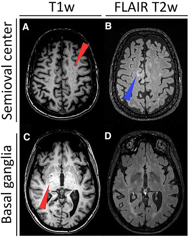
Perivascular spaces can become detectable on magnetic resonance imaging (MRI) upon enlargement, referred to as enlarged perivascular spaces (EPVS) or Virchow-Robin spaces. EPVS have been linked to small vessel disease. Some studies have also indicated an association of EPVS to neuroinflammation and/or neurodegeneration. However, there is conflicting evidence with regards to their potential as a clinically relevant imaging biomarker in multiple sclerosis (MS).

METHODS

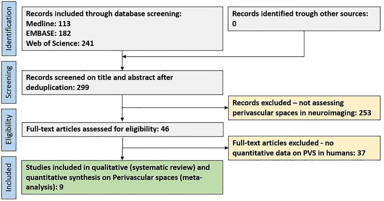
To perform a systematic review and meta-analysis of EPVS as visualized by MRI in MS. Nine out of 299 original studies addressing EPVS in humans using MRI were eligible for the systematic review and meta-analysis including a total of 457 MS patients and 352 control subjects.

RESULTS

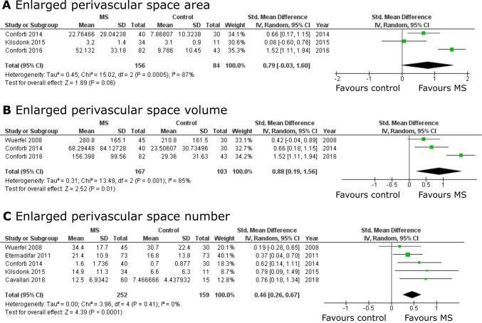
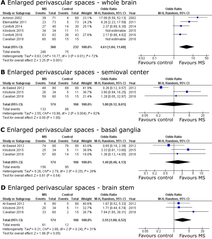
In MS, EPVS have been associated with cognitive decline, contrast-enhancing MRI lesions, and brain atrophy. Yet, these associations were not consistent between studies. The meta-analysis revealed that MS patients have greater EPVS prevalence (odds ratio = 4.61, 95% CI = [1.84; 11.60], p = 0.001) as well as higher EPVS counts (standardized mean difference [SMD] = 0.46, 95% CI = [0.26; 0.67], p < 0.001) and larger volumes (SMD = 0.88, 95% CI = [0.19; 1.56], p = 0.01) compared to controls.

CONCLUSIONS

Available literature suggests a higher EPVS burden in MS patients compared to controls. The association of EPVS to neuroinflammatory or -degenerative pathology in MS remains inconsistent. Thus, there is currently insufficient evidence supporting EPVS as diagnostic and/or prognostic marker in MS. In order to benefit future comparisons of studies, we propose recommendations on EPVS assessment standardization in MS. PROSPERO No: CRD42019133946.

摘要

背景

血管周围间隙在扩大时可在磁共振成像(MRI)上检测到,称为扩大的血管周围间隙(EPVS)或 Virchow-Robin 空间。EPVS 与小血管疾病有关。一些研究还表明 EPVS 与神经炎症和/或神经退行性变有关。然而,在多发性硬化症(MS)中,EPVS 作为一种有临床意义的影像学生物标志物的潜力存在相互矛盾的证据。

方法

对 MRI 显示的 MS 中 EPVS 进行系统评价和荟萃分析。9 项共 299 项原始研究中,有 9 项使用 MRI 研究人类 EPVS,符合系统评价和荟萃分析的标准,共纳入 457 例 MS 患者和 352 例对照。

结果

在 MS 中,EPVS 与认知能力下降、对比增强 MRI 病变和脑萎缩有关。然而,这些关联在研究之间并不一致。荟萃分析显示,MS 患者的 EPVS 患病率更高(优势比=4.61,95%可信区间[1.84;11.60],p=0.001),EPVS 计数更高(标准化均数差[SMD]=0.46,95%可信区间[0.26;0.67],p<0.001)和更大的体积(SMD=0.88,95%可信区间[0.19;1.56],p=0.01)与对照组相比。

结论

现有文献表明,MS 患者的 EPVS 负担高于对照组。EPVS 与 MS 中的神经炎症或神经退行性病变的关联仍然不一致。因此,目前没有足够的证据支持 EPVS 作为 MS 的诊断和/或预后标志物。为了使未来的研究比较受益,我们提出了 MS 中 EPVS 评估标准化的建议。PROSPERO 编号:CRD42019133946。

https://cdn.ncbi.nlm.nih.gov/pmc/blobs/67b2/7577911/4b5a34823857/415_2020_9971_Fig4_HTML.jpg
https://cdn.ncbi.nlm.nih.gov/pmc/blobs/67b2/7577911/897426afdae7/415_2020_9971_Fig1_HTML.jpg
https://cdn.ncbi.nlm.nih.gov/pmc/blobs/67b2/7577911/5a3840f4b675/415_2020_9971_Fig2_HTML.jpg
https://cdn.ncbi.nlm.nih.gov/pmc/blobs/67b2/7577911/06dcceff9cc3/415_2020_9971_Fig3_HTML.jpg
https://cdn.ncbi.nlm.nih.gov/pmc/blobs/67b2/7577911/4b5a34823857/415_2020_9971_Fig4_HTML.jpg

相似文献

1
Enlarged perivascular spaces in multiple sclerosis on magnetic resonance imaging: a systematic review and meta-analysis.磁共振成像多发性硬化症中的血管周围间隙扩大:系统评价和荟萃分析。
J Neurol. 2020 Nov;267(11):3199-3212. doi: 10.1007/s00415-020-09971-5. Epub 2020 Jun 13.
2
Enlarged perivascular spaces are associated with brain microangiopathy and aging in multiple sclerosis.扩大的血管周围间隙与多发性硬化症中的脑微血管病和衰老有关。
Mult Scler. 2024 Jul;30(8):983-993. doi: 10.1177/13524585241256881. Epub 2024 Jun 7.
3
Enlarged Perivascular Spaces and Dementia: A Systematic Review.血管周围间隙扩大与痴呆:系统综述。
J Alzheimers Dis. 2019;72(1):247-256. doi: 10.3233/JAD-190527.
4
Association of Enlarged Perivascular Spaces and Measures of Small Vessel and Alzheimer Disease.血管周围间隙扩大与小血管和阿尔茨海默病相关因素的分析。
Neurology. 2021 Jan 12;96(2):e193-e202. doi: 10.1212/WNL.0000000000011046. Epub 2020 Oct 12.
5
Evaluating the Association between Enlarged Perivascular Spaces and Disease Worsening in Multiple Sclerosis.评估多发性硬化症中增大的血管周围空间与疾病恶化之间的关联。
J Neuroimaging. 2018 May;28(3):273-277. doi: 10.1111/jon.12490. Epub 2017 Dec 11.
6
Enlarged Perivascular Spaces Are Independently Associated with High Pulse Wave Velocity: A Cross-Sectional Study.血管周围空间扩大与脉搏波速度升高独立相关:一项横断面研究。
J Alzheimers Dis. 2024;101(2):627-636. doi: 10.3233/JAD-240589.
7
The etiology and evolution of magnetic resonance imaging-visible perivascular spaces: Systematic review and meta-analysis.磁共振成像可见的血管周围间隙的病因及演变:系统评价与荟萃分析
Front Neurosci. 2023 Mar 30;17:1038011. doi: 10.3389/fnins.2023.1038011. eCollection 2023.
8
The relationship between blood-brain barrier permeability and enlarged perivascular spaces: a cross-sectional study.血脑屏障通透性与扩大的血管周围间隙之间的关系:一项横断面研究。
Clin Interv Aging. 2019 May 10;14:871-878. doi: 10.2147/CIA.S204269. eCollection 2019.
9
Associations of Enlarged Perivascular Spaces With Brain Lesions, Brain Age, and Clinical Outcomes in Chronic Traumatic Brain Injury.血管周围空间扩大与脑损伤、脑龄和慢性创伤性脑损伤临床结局的关联。
Neurology. 2023 Jul 4;101(1):e63-e73. doi: 10.1212/WNL.0000000000207370. Epub 2023 May 8.
10
Neuropathologic and Cognitive Correlates of Enlarged Perivascular Spaces in a Community-Based Cohort of Older Adults.社区老年人队列中血管周围间隙扩大的神经病理学和认知相关性研究。
Stroke. 2020 Sep;51(9):2825-2833. doi: 10.1161/STROKEAHA.120.029388. Epub 2020 Aug 6.

引用本文的文献

1
Sleep and Risk of Multiple Sclerosis: Bridging the Gap Between Inflammation and Neurodegeneration via Glymphatic Failure.睡眠与多发性硬化症风险:通过类淋巴系统功能障碍弥合炎症与神经退行性变之间的差距
Brain Sci. 2025 Jul 19;15(7):766. doi: 10.3390/brainsci15070766.
2
Cognitive Brain Networks and Enlarged Perivascular Spaces: Implications for Symptom Severity and Support Needs in Children with Autism.认知脑网络与血管周围间隙增宽:对自闭症儿童症状严重程度及支持需求的影响
J Clin Med. 2025 Apr 27;14(9):3029. doi: 10.3390/jcm14093029.
3
Enlarged perivascular spaces under the dorso-lateral prefrontal cortex and severity of autism.

本文引用的文献

1
Perivascular spaces in the brain: anatomy, physiology and pathology.脑内血管周围间隙:解剖、生理与病理。
Nat Rev Neurol. 2020 Mar;16(3):137-153. doi: 10.1038/s41582-020-0312-z. Epub 2020 Feb 24.
2
High-degree centrum semiovale-perivascular spaces are associated with development of subdural fluid in mild traumatic brain injury.高等级半卵圆中心-血管周围间隙与轻度外伤性脑损伤硬膜下积液的发展相关。
PLoS One. 2019 Sep 4;14(9):e0221788. doi: 10.1371/journal.pone.0221788. eCollection 2019.
3
Autoidentification of perivascular spaces in white matter using clinical field strength T and FLAIR MR imaging.
背外侧前额叶皮质下血管周围间隙扩大与自闭症严重程度
Sci Rep. 2025 Mar 9;15(1):8142. doi: 10.1038/s41598-025-92913-w.
4
MRI quantified enlarged perivascular space volumes as imaging biomarkers correlating with severity of anxiety depression in young adults with long-time mobile phone use.磁共振成像(MRI)将扩大的血管周围间隙体积量化为成像生物标志物,这些标志物与长期使用手机的年轻人的焦虑抑郁严重程度相关。
Front Psychiatry. 2025 Feb 20;16:1532256. doi: 10.3389/fpsyt.2025.1532256. eCollection 2025.
5
A Novel Mouse Model for Cerebral Inflammatory Demyelination in X-Linked Adrenoleukodystrophy: Insights into Pathogenesis and Potential Therapeutic Targets.一种用于X连锁肾上腺脑白质营养不良症脑炎性脱髓鞘的新型小鼠模型:对发病机制和潜在治疗靶点的见解
Ann Neurol. 2025 Feb;97(2):296-312. doi: 10.1002/ana.27117. Epub 2024 Oct 28.
6
Glymphatic dysfunction in multiple sclerosis and its association with disease pathology and disability.多发性硬化症中的糖质分解代谢功能障碍及其与疾病病理和残疾的关系。
Mult Scler. 2024 Nov;30(13):1609-1619. doi: 10.1177/13524585241280842. Epub 2024 Sep 29.
7
Focally Enlarged Perivascular Spaces in Pediatric and Adolescent Patients with Polymicrogyria-an MRI Study.多小脑回畸形的儿童和青少年患者中局部扩大的血管周围间隙——一项MRI研究
Clin Neuroradiol. 2025 Mar;35(1):87-93. doi: 10.1007/s00062-024-01457-5. Epub 2024 Sep 13.
8
Paramagnetic rim lesions as a biomarker to discriminate between multiple sclerosis and cerebral small vessel disease.顺磁性边缘病变作为区分多发性硬化症和脑小血管疾病的生物标志物
Front Neurol. 2024 Jul 16;15:1429698. doi: 10.3389/fneur.2024.1429698. eCollection 2024.
9
Changes of brain parenchyma free water fraction reflect tissue damage and impaired processing speed in multiple sclerosis.脑实质自由水分数的变化反映了多发性硬化中的组织损伤和处理速度受损。
Hum Brain Mapp. 2024 Jun 15;45(9):e26761. doi: 10.1002/hbm.26761.
10
Glymphatic system: a gateway for neuroinflammation.类淋巴系统:神经炎症的一个通道。
Neural Regen Res. 2024 Dec 1;19(12):2661-2672. doi: 10.4103/1673-5374.391312. Epub 2023 Dec 21.
使用临床场强 T 和 FLAIR MR 成像自动识别脑白质血管周围间隙。
Neuroimage. 2019 Nov 15;202:116126. doi: 10.1016/j.neuroimage.2019.116126. Epub 2019 Aug 25.
4
Perivascular spaces and their associations with risk factors, clinical disorders and neuroimaging features: A systematic review and meta-analysis.血管周围间隙及其与危险因素、临床疾病和神经影像学特征的关系:系统评价和荟萃分析。
Int J Stroke. 2019 Jun;14(4):359-371. doi: 10.1177/1747493019830321. Epub 2019 Feb 14.
5
Blood-brain barrier breakdown is an early biomarker of human cognitive dysfunction.血脑屏障破坏是人类认知功能障碍的早期生物标志物。
Nat Med. 2019 Feb;25(2):270-276. doi: 10.1038/s41591-018-0297-y. Epub 2019 Jan 14.
6
Clinical Significance of Magnetic Resonance Imaging Markers of Vascular Brain Injury: A Systematic Review and Meta-analysis.磁共振血管损伤标志物的临床意义:系统评价和荟萃分析。
JAMA Neurol. 2019 Jan 1;76(1):81-94. doi: 10.1001/jamaneurol.2018.3122.
7
Enlarged perivascular spaces in brain MRI: Automated quantification in four regions.脑 MRI 中的血管周围间隙扩大:四个区域的自动量化。
Neuroimage. 2019 Jan 15;185:534-544. doi: 10.1016/j.neuroimage.2018.10.026. Epub 2018 Oct 13.
8
Repeatability and reproducibility of FreeSurfer, FSL-SIENAX and SPM brain volumetric measurements and the effect of lesion filling in multiple sclerosis.FreeSurfer、FSL-SIENAX 和 SPM 脑容量测量的可重复性和再现性,以及多发性硬化症中病变填充的影响。
Eur Radiol. 2019 Mar;29(3):1355-1364. doi: 10.1007/s00330-018-5710-x. Epub 2018 Sep 21.
9
Is bulk flow plausible in perivascular, paravascular and paravenous channels?在血管周隙、旁血管和旁静脉通道中,整体流动是否合理?
Fluids Barriers CNS. 2018 Jun 15;15(1):17. doi: 10.1186/s12987-018-0103-8.
10
The fine anatomy of the perivascular compartment in the human brain: relevance to dilated perivascular spaces in cerebral amyloid angiopathy.人脑血管周围间隙的精细解剖结构:与脑淀粉样血管病中血管周围间隙扩张的相关性
Neuropathol Appl Neurobiol. 2019 Apr;45(3):305-308. doi: 10.1111/nan.12480. Epub 2018 May 15.