Department of Medical Oncology, Faculty of Medicine, Gazi University, Ankara, Turkey.
Department of Pathology, Faculty of Medicine, Gazi University, Ankara, Turkey.
Turk J Med Sci. 2023 Feb;53(1):142-148. doi: 10.55730/1300-0144.5567. Epub 2023 Feb 22.
This study aimed to evaluate the expression of lymphocyte activation gene-3 (LAG-3) and its relationship with programmed cell death ligand-1 (PD-L1) in triple-negative breast cancer (TNBC).
: LAG-3 and PD-L1 was evaluated in tumor-infiltrating lymphocytes (TILs) using immunohistochemistry (IHC). The chi-square test was used to estimate the associations between LAG-3, PD-L1 and clinicopathological characteristics. Correlation between LAG-3 stromal TIL (sTIL), LAG-3 intraepitelial TIL (iTIL) and PD-L1 was assessed with using the Spearman's correlation coefficient. Survival analysis was performed using the Kaplan-Meier method.
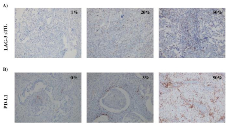
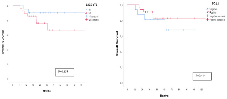
The percentages of LAG-3 sTIL+, LAG-3 iTIL+, PD-L1+ tumor cells and PD-L1+ inflammatory cells were 52%, 42%, 14% and 70%, respectively. A strong positive correlation between LAG-3 sTIL and LAG-3 iTIL (r = 0.874, p < 0.001) and a moderate positive correlation between LAG-3 sTIL and PD-L1 (r = 0.584, p < 0.001) were found. LAG-3 and PD-L1 status did not significantly affect overall survival (OS) (HR: 0.56 (95% CI: 0.15-2.11) (p = 0.397), HR: 2.70 (95% CI: 0.56-13.02) (p = 0.215), respectively).
High levels of LAG-3 and PD-L1 expression were detected in patients with TNBC. Although their contribution to survival could not be determined, the high expression rates of PD-L1 and LAG-3 may help identify the subgroup of TNBC that would benefit from immunotherapy.
本研究旨在评估淋巴细胞激活基因 3(LAG-3)的表达及其与程序性死亡配体 1(PD-L1)在三阴性乳腺癌(TNBC)中的关系。
使用免疫组织化学(IHC)评估肿瘤浸润淋巴细胞(TIL)中的 LAG-3 和 PD-L1。使用卡方检验估计 LAG-3、PD-L1 与临床病理特征之间的关联。使用 Spearman 相关系数评估 LAG-3 基质 TIL(sTIL)、LAG-3 上皮内 TIL(iTIL)与 PD-L1 之间的相关性。使用 Kaplan-Meier 方法进行生存分析。
LAG-3 sTIL+、LAG-3 iTIL+、PD-L1+肿瘤细胞和 PD-L1+炎症细胞的百分比分别为 52%、42%、14%和 70%。LAG-3 sTIL 与 LAG-3 iTIL 之间存在强烈的正相关(r = 0.874,p < 0.001),LAG-3 sTIL 与 PD-L1 之间存在中度正相关(r = 0.584,p < 0.001)。LAG-3 和 PD-L1 状态对总生存(OS)没有显著影响(HR:0.56(95%CI:0.15-2.11)(p = 0.397),HR:2.70(95%CI:0.56-13.02)(p = 0.215))。
在 TNBC 患者中检测到高水平的 LAG-3 和 PD-L1 表达。尽管无法确定它们对生存的贡献,但 PD-L1 和 LAG-3 的高表达率可能有助于确定从免疫治疗中受益的 TNBC 亚组。