Department of Orthopaedics, The Second Hospital, Cheeloo College of Medicine, Shandong University, 247 Beiyuan Street, Jinan, 250033, Shandong, People's Republic of China.
J Orthop Surg Res. 2023 Mar 27;18(1):248. doi: 10.1186/s13018-023-03717-5.
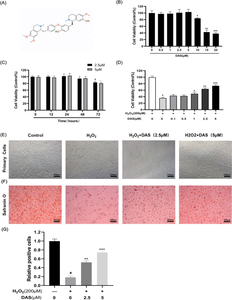
Osteoarthritis (OA) is a chronic degenerative joint disease characterized by cartilage degeneration and intra-articular inflammation. Daurisoline (DAS) is an isoquinoline alkaloid isolated from Rhizoma Menispermi, whose antitumor and anti-inflammatory pharmacological effects have been demonstrated, but the effects of DAS on OA have rarely been researched. In this study, we aimed to explore the potential role of DAS in OA and its partial mechanism.
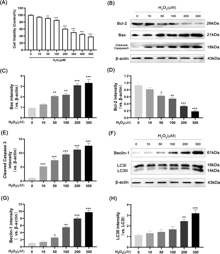
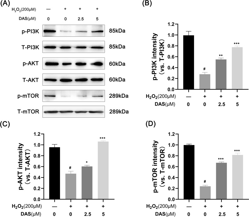
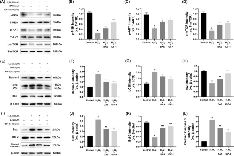
The cytotoxicity of HO and DAS toward chondrocytes was detected by the Cell Counting Kit-8 assay. Safranin O staining was used to detect chondrocyte phenotype changes. Cell apoptosis was measured by both flow cytometry and quantitative analysis of the protein levels of the apoptosis-related factors Bax, Bcl-2 and cleaved caspase-3 by western blot. Western blotting and immunofluorescence were used to assess the expression of the autophagy-related proteins LC3, Beclin-1 and p62. In addition, key signal pathway targets and matrix-degrading indicators were measured by western blot.
Our results indicated that HO induced human chondrocyte apoptosis and activated autophagy in a dose-dependent manner. DAS treatment dose-dependently reversed the expression of apoptosis-related proteins (Bax, Bcl-2 and cleaved caspase3) and the apoptosis rate induced by HO. Western blot and immunofluorescence analyses showed that DAS decreased the HO-induced upregulation of the autophagy marker Beclin-1 and the LC3 II/LC3 I ratio and upregulated the p62 protein level. Mechanistically, DAS inhibited autophagy through the activation of the classical PI3K/AKT/mTOR signaling pathway and protected chondrocytes from apoptosis. In addition, DAS alleviated the HO-induced degradation of type II collagen and the high expression of matrix metalloproteinase 3 (MMP3) and MMP13.
Our research demonstrated that DAS alleviated chondrocyte autophagy caused by HO through activation of the PI3K/AKT/mTOR signaling pathway and protected chondrocytes from apoptosis and matrix degradation. In conclusion, these findings suggest that DAS may serve as a promising therapeutic strategy for OA.
骨关节炎(OA)是一种慢性退行性关节疾病,其特征为软骨退化和关节内炎症。蝙蝠葛苏林碱(DAS)是从防己科蝙蝠葛属植物蝙蝠葛根茎中分离得到的一种异喹啉类生物碱,其具有抗肿瘤和抗炎的药理学作用,但 DAS 对 OA 的作用鲜有研究。在本研究中,我们旨在探讨 DAS 在 OA 中的潜在作用及其部分机制。
通过细胞计数试剂盒-8 检测 HO 和 DAS 对软骨细胞的细胞毒性。通过番红 O 染色检测软骨细胞表型变化。通过流式细胞术和蛋白质印迹法对凋亡相关因子 Bax、Bcl-2 和 cleaved caspase-3 的蛋白水平进行定量分析来测量细胞凋亡。通过蛋白质印迹法和免疫荧光法评估自噬相关蛋白 LC3、Beclin-1 和 p62 的表达。此外,通过蛋白质印迹法测量关键信号通路靶点和基质降解指标。
我们的结果表明,HO 以剂量依赖性方式诱导人软骨细胞凋亡并激活自噬。DAS 处理剂量依赖性地逆转了 HO 诱导的凋亡相关蛋白(Bax、Bcl-2 和 cleaved caspase3)表达和凋亡率。Western blot 和免疫荧光分析表明,DAS 降低了 HO 诱导的自噬标志物 Beclin-1 和 LC3 II/LC3 I 比值的上调,并上调了 p62 蛋白水平。机制上,DAS 通过激活经典的 PI3K/AKT/mTOR 信号通路抑制自噬并保护软骨细胞免于凋亡。此外,DAS 减轻了 HO 诱导的 II 型胶原降解以及基质金属蛋白酶 3(MMP3)和基质金属蛋白酶 13(MMP13)的高表达。
我们的研究表明,DAS 通过激活 PI3K/AKT/mTOR 信号通路减轻 HO 诱导的软骨细胞自噬,从而保护软骨细胞免于凋亡和基质降解。总之,这些发现表明 DAS 可能是 OA 的一种有前途的治疗策略。