Department of Biomedical Engineering, Texas A&M University, College Station, Texas, United States of America.
Microscopy and Imaging Center, Texas A&M University, College Station, Texas, United States of America.
PLoS One. 2023 Mar 28;18(3):e0282298. doi: 10.1371/journal.pone.0282298. eCollection 2023.
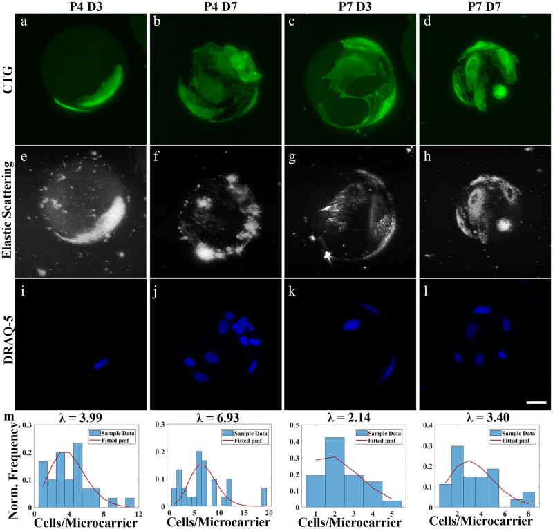
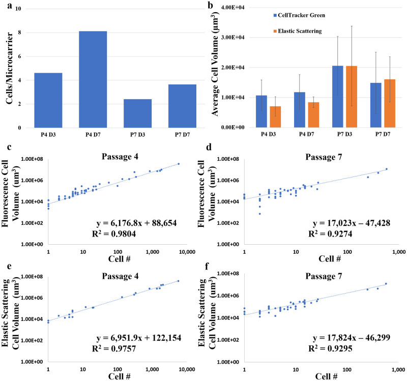
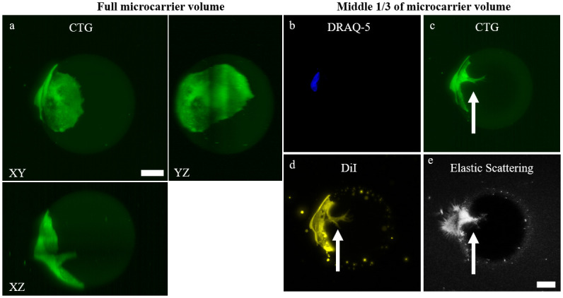
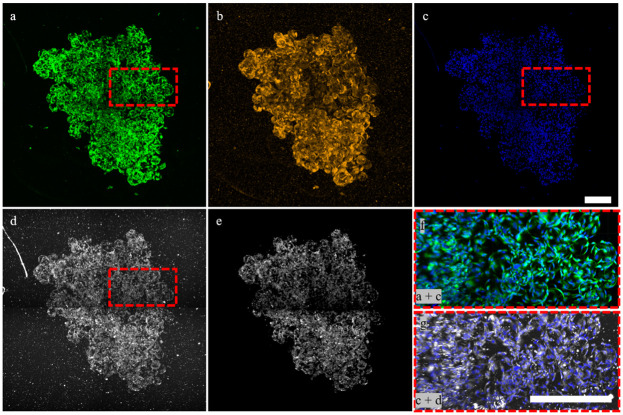
The adoption of cell-based therapies into the clinic will require tremendous large-scale expansion to satisfy future demand, and bioreactor-microcarrier cultures are best suited to meet this challenge. The use of spherical microcarriers, however, precludes in-process visualization and monitoring of cell number, morphology, and culture health. The development of novel expansion methods also motivates the advancement of analytical methods used to characterize these microcarrier cultures. A robust optical imaging and image-analysis assay to non-destructively quantify cell number and cell volume was developed. This method preserves 3D cell morphology and does not require membrane lysing, cellular detachment, or exogenous labeling. Complex cellular networks formed in microcarrier aggregates were imaged and analyzed in toto. Direct cell enumeration of large aggregates was performed in toto for the first time. This assay was successfully applied to monitor cellular growth of mesenchymal stem cells attached to spherical hydrogel microcarriers over time. Elastic scattering and fluorescence lightsheet microscopy were used to quantify cell volume and cell number at varying spatial scales. The presented study motivates the development of on-line optical imaging and image analysis systems for robust, automated, and non-destructive monitoring of bioreactor-microcarrier cell cultures.
细胞疗法的临床应用需要进行大规模的扩增,以满足未来的需求,而生物反应器-微载体培养是最适合应对这一挑战的方法。然而,使用球形微载体则无法在过程中对细胞数量、形态和培养物健康进行可视化和监测。新型扩增方法的发展也促使人们开发用于表征这些微载体培养物的分析方法。本文开发了一种强大的光学成像和图像分析测定法,可无损地定量细胞数量和细胞体积。该方法保留了 3D 细胞形态,不需要膜裂解、细胞分离或外源标记。对微载体聚集体中形成的复杂细胞网络进行了整体成像和分析。首次对大聚集体进行了直接细胞计数。该测定法成功地应用于监测附着在球形水凝胶微载体上的间充质干细胞随时间的细胞生长。弹性散射和荧光光片显微镜被用于在不同的空间尺度上定量细胞体积和细胞数量。本研究促进了在线光学成像和图像分析系统的开发,用于对生物反应器-微载体细胞培养物进行稳健、自动化和无损监测。