Texas A&M University, Department of Biomedical Engineering, College Station, Texas, United States.
Texas A&M University, Microscopy and Imaging Center, College Station, Texas, United States.
J Biomed Opt. 2024 Jun;29(Suppl 2):S22708. doi: 10.1117/1.JBO.29.S2.S22708. Epub 2024 Jun 13.
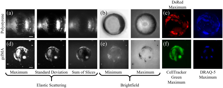
The ability to observe and monitor cell density and morphology has been imperative for assessing the health of a cell culture and for producing high quality, high yield cell cultures for decades. Microcarrier-based cultures, used for large-scale cellular expansion processes, are not compatible with traditional visualization-based methods, such as widefield microscopy, due to their thickness and material composition.
Here, we assess the optical imaging compatibilities of commercial polystyrene microcarriers versus custom-fabricated gelatin methacryloyl (gelMA) microcarriers for non-destructive and non-invasive visualization of the entire microcarrier surface, direct cell enumeration, and sub-cellular visualization of mesenchymal stem/stromal cells.
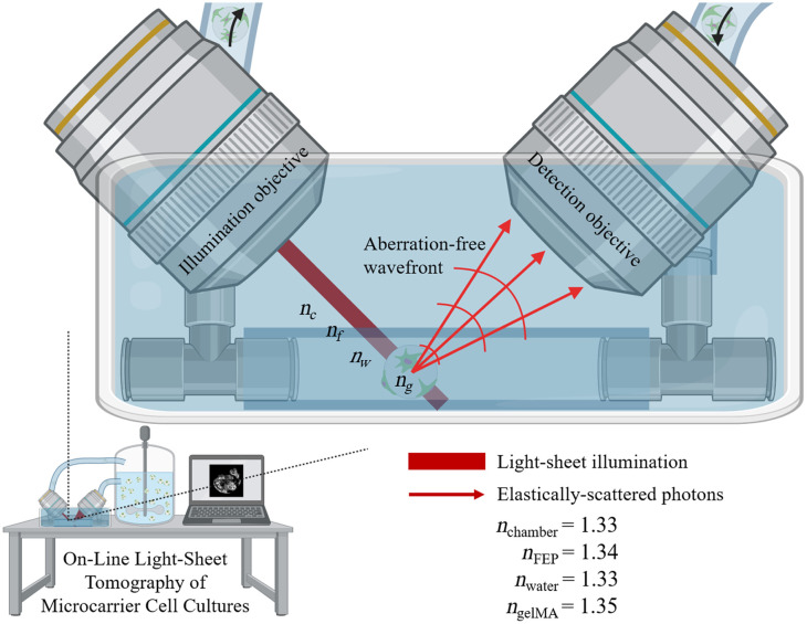
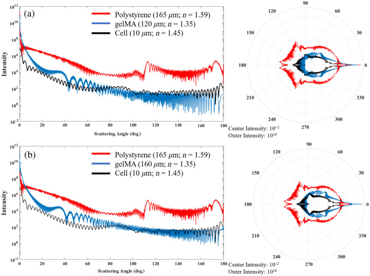
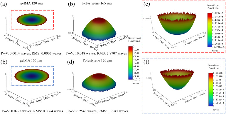
Mie scattering and wavefront error simulations of the polystyrene and gelMA microcarriers were performed to assess the potential for elastic scattering-based imaging of adherent cells. A Zeiss Z.1 light-sheet microscope was adapted to perform light-sheet tomography using label-free elastic scattering contrast from planar side illumination to achieve optical sectioning and permit non-invasive and non-destructive, , three-dimensional, high-resolution visualization of cells cultured on microcarriers.
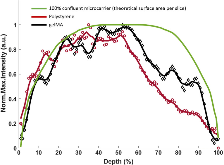
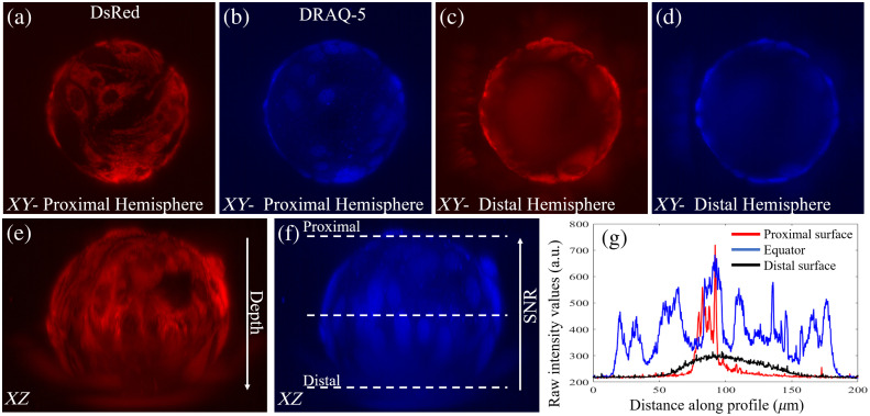
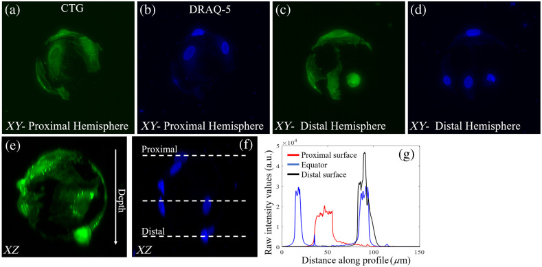
The polystyrene microcarrier prevents visualization of cells on the distal half of the microcarrier using either fluorescence or elastic scattering contrast, whereas the gelMA microcarrier allows for high fidelity visualization of cell morphology and quantification of cell density using light-sheet fluorescence microscopy and tomography.
The combination of optical-quality gelMA microcarriers and label-free light-sheet tomography will facilitate enhanced control of bioreactor-microcarrier cell culture processes.
几十年来,观察和监测细胞密度和形态一直是评估细胞培养物健康状况和生产高质量、高产细胞培养物的必要条件。由于其厚度和材料组成,基于微载体的培养物不适合用于大规模细胞扩展过程的传统基于可视化的方法,例如宽场显微镜。
在这里,我们评估了商业聚苯乙烯微载体与定制明胶甲基丙烯酰(gelMA)微载体在非破坏性和非侵入性可视化整个微载体表面、直接细胞计数以及间充质干细胞的亚细胞可视化方面的光学成像兼容性。
对聚苯乙烯和 gelMA 微载体进行 Mie 散射和波前误差模拟,以评估基于弹性散射对贴壁细胞成像的潜力。对 Zeiss Z.1 光片显微镜进行了改造,使用来自平面侧照明的无标记弹性散射对比进行光片断层扫描,以实现光学切片,并允许对微载体上培养的细胞进行非侵入性和非破坏性的、三维的、高分辨率可视化。
聚苯乙烯微载体使用荧光或弹性散射对比都阻止了对微载体远端一半上细胞的可视化,而 gelMA 微载体允许使用光片荧光显微镜和断层扫描对细胞形态进行高保真可视化和细胞密度的定量。
光学质量的 gelMA 微载体和无标记光片断层扫描的结合将促进对生物反应器-微载体细胞培养过程的增强控制。