• 文献检索
  • 文档翻译
  • 深度研究
  • 学术资讯
  • Suppr Zotero 插件Zotero 插件
  • 邀请有礼
  • 套餐&价格
  • 历史记录
应用&插件
Suppr Zotero 插件Zotero 插件浏览器插件Mac 客户端Windows 客户端微信小程序
定价
高级版会员购买积分包购买API积分包
服务
文献检索文档翻译深度研究API 文档MCP 服务
关于我们
关于 Suppr公司介绍联系我们用户协议隐私条款
关注我们

Suppr 超能文献

核心技术专利:CN118964589B侵权必究
粤ICP备2023148730 号-1Suppr @ 2026

文献检索

告别复杂PubMed语法,用中文像聊天一样搜索,搜遍4000万医学文献。AI智能推荐,让科研检索更轻松。

立即免费搜索

文件翻译

保留排版,准确专业,支持PDF/Word/PPT等文件格式,支持 12+语言互译。

免费翻译文档

深度研究

AI帮你快速写综述,25分钟生成高质量综述,智能提取关键信息,辅助科研写作。

立即免费体验

创伤性脑损伤动物模型的建立:局部损伤后异位骨化的增强

Development of an Animal Model for Traumatic Brain Injury Augmentation of Heterotopic Ossification in Response to Local Injury.

作者信息

Kesavan Chandrasekhar, Gomez Gustavo A, Pourteymoor Sheila, Mohan Subburaman

机构信息

Musculoskeletal Disease Center, VA Loma Linda Healthcare System, Loma Linda, CA 92357, USA.

Department of Medicine, Loma Linda University, Loma Linda, CA 92354, USA.

出版信息

Biomedicines. 2023 Mar 18;11(3):943. doi: 10.3390/biomedicines11030943.

DOI:10.3390/biomedicines11030943
PMID:36979922
原文链接:https://pmc.ncbi.nlm.nih.gov/articles/PMC10046150/
Abstract

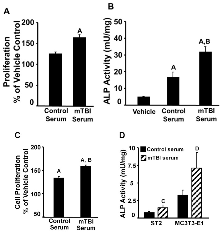
Heterotopic ossification (HO) is the abnormal growth of bone in soft connective tissues that occurs as a frequent complication in individuals with traumatic brain injury (TBI) and in rare genetic disorders. Therefore, understanding the mechanisms behind ectopic bone formation in response to TBI is likely to have a significant impact on identification of novel therapeutic targets for HO treatment. In this study, we induced repetitive mild TBI (mTBI) using a weight drop model in mice and then stimulated HO formation via a local injury to the Achilles tendon or fibula. The amount of ectopic bone, as evaluated by micro-CT analyses, was increased by four-fold in the injured leg of mTBI mice compared to control mice. However, there was no evidence of HO formation in the uninjured leg of mTBI mice. Since tissue injury leads to the activation of hypoxia signaling, which is known to promote endochondral ossification, we evaluated the effect of IOX2, a chemical inhibitor of PHD2 and a known inducer of hypoxia signaling on HO development in response to fibular injury. IOX2 treatment increased HO volume by five-fold compared to vehicle. Since pericytes located in the endothelium of microvascular capillaries are known to function as multipotent tissue-resident progenitors, we determined if activation of hypoxia signaling promotes pericyte recruitment at the injury site. We found that markers of pericytes, NG2 and PDGFRβ, were abundantly expressed at the site of injury in IOX2 treated mice. Treatment of pericytes with IOX2 for 72 h stimulated expression of targets of hypoxia signaling ( and ), as well as markers of chondrocyte differentiation ( and ). Furthermore, serum collected from TBI mice was more effective in promoting the proliferation and differentiation of pericytes than control mouse serum. In conclusion, our data show that the hypoxic state at the injury site in soft tissues of TBI mice provides an environment leading to increased accumulation and activation of pericytes to form endochondral bone.

摘要

异位骨化(HO)是指在软结缔组织中发生的异常骨生长,它是创伤性脑损伤(TBI)患者常见的并发症,在罕见的遗传疾病中也会出现。因此,了解TBI后异位骨形成的机制可能对确定HO治疗的新靶点产生重大影响。在本研究中,我们使用重物坠落模型在小鼠中诱导重复性轻度TBI(mTBI),然后通过对跟腱或腓骨的局部损伤刺激HO形成。通过微计算机断层扫描(micro-CT)分析评估,与对照小鼠相比,mTBI小鼠受伤腿部的异位骨量增加了四倍。然而,在mTBI小鼠未受伤的腿部没有HO形成的证据。由于组织损伤会导致缺氧信号的激活,而缺氧信号已知会促进软骨内骨化,我们评估了IOX2(一种PHD2的化学抑制剂和已知的缺氧信号诱导剂)对腓骨损伤后HO发展的影响。与载体相比,IOX2处理使HO体积增加了五倍。由于位于微血管内皮的周细胞已知具有多能组织驻留祖细胞的功能,我们确定缺氧信号的激活是否会促进损伤部位的周细胞募集。我们发现,在IOX2处理的小鼠的损伤部位,周细胞标志物NG2和血小板衍生生长因子受体β(PDGFRβ)大量表达。用IOX2处理周细胞72小时可刺激缺氧信号靶点(和)以及软骨细胞分化标志物(和)的表达。此外,从TBI小鼠收集的血清在促进周细胞增殖和分化方面比对照小鼠血清更有效。总之,我们的数据表明,TBI小鼠软组织损伤部位的缺氧状态提供了一个环境,导致周细胞的积累和激活增加,从而形成软骨内骨。

https://cdn.ncbi.nlm.nih.gov/pmc/blobs/08c1/10046150/d6f56270872e/biomedicines-11-00943-g004.jpg
https://cdn.ncbi.nlm.nih.gov/pmc/blobs/08c1/10046150/f46fdd9b5672/biomedicines-11-00943-g001.jpg
https://cdn.ncbi.nlm.nih.gov/pmc/blobs/08c1/10046150/9ee9beaabcbb/biomedicines-11-00943-g002.jpg
https://cdn.ncbi.nlm.nih.gov/pmc/blobs/08c1/10046150/16cd1e5b04b4/biomedicines-11-00943-g003.jpg
https://cdn.ncbi.nlm.nih.gov/pmc/blobs/08c1/10046150/d6f56270872e/biomedicines-11-00943-g004.jpg

相似文献

1
Development of an Animal Model for Traumatic Brain Injury Augmentation of Heterotopic Ossification in Response to Local Injury.创伤性脑损伤动物模型的建立:局部损伤后异位骨化的增强
Biomedicines. 2023 Mar 18;11(3):943. doi: 10.3390/biomedicines11030943.
2
Relationship between heterotopic ossification and traumatic brain injury: Why severe traumatic brain injury increases the risk of heterotopic ossification.异位骨化与创伤性脑损伤之间的关系:为何重度创伤性脑损伤会增加异位骨化的风险。
J Orthop Translat. 2017 Nov 14;12:16-25. doi: 10.1016/j.jot.2017.10.002. eCollection 2018 Jan.
3
mTORC1 coordinates NF-κB signaling pathway to promote chondrogenic differentiation of tendon cells in heterotopic ossification.mTORC1 协调 NF-κB 信号通路促进异位骨化中肌腱细胞的软骨分化。
Bone. 2022 Oct;163:116507. doi: 10.1016/j.bone.2022.116507. Epub 2022 Jul 29.
4
Early detection of burn induced heterotopic ossification using transcutaneous Raman spectroscopy.利用经皮拉曼光谱术早期检测烧伤诱导性异位骨化。
Bone. 2013 May;54(1):28-34. doi: 10.1016/j.bone.2013.01.002. Epub 2013 Jan 11.
5
Small molecule inhibition of non-canonical (TAK1-mediated) BMP signaling results in reduced chondrogenic ossification and heterotopic ossification in a rat model of blast-associated combat-related lower limb trauma.在爆炸相关战斗所致下肢创伤的大鼠模型中,小分子抑制非经典(TAK1介导)骨形态发生蛋白信号传导可导致软骨内成骨及异位骨化减少。
Bone. 2020 Oct;139:115517. doi: 10.1016/j.bone.2020.115517. Epub 2020 Jul 2.
6
The Impact of Cigarette Smoking on the Formation of Heterotopic Ossification Among Service Members With a Traumatic Amputation.吸烟对创伤性截肢军人异位骨化形成的影响。
Mil Med. 2017 May;182(5):e1742-e1748. doi: 10.7205/MILMED-D-16-00350.
7
βig-h3 enhances chondrogenesis via promoting mesenchymal condensation in rat Achilles tendon heterotopic ossification model.βig-h3 通过促进大鼠跟腱异位骨化模型中成纤维细胞凝聚促进软骨生成。
Aging (Albany NY). 2020 Apr 20;12(8):7030-7041. doi: 10.18632/aging.103060.
8
Traumatic brain injury enhances the formation of heterotopic ossification around the hip: an animal model study.创伤性脑损伤增强了髋周异位骨化的形成:一项动物模型研究。
Arch Orthop Trauma Surg. 2020 Aug;140(8):1029-1035. doi: 10.1007/s00402-019-03326-0. Epub 2019 Dec 13.
9
Echinomycin in the prevention of heterotopic ossification - an experimental antibiotic agent shows promising results in a murine model.依诺霉素预防异位骨化 - 一种实验性抗生素在小鼠模型中显示出良好的效果。
Injury. 2013 Apr;44(4):570-5. doi: 10.1016/j.injury.2012.12.030. Epub 2013 Feb 8.
10
Analysis of Bone-Cartilage-Stromal Progenitor Populations in Trauma Induced and Genetic Models of Heterotopic Ossification.创伤诱导和异位骨化遗传模型中骨-软骨-基质祖细胞群体的分析
Stem Cells. 2016 Jun;34(6):1692-701. doi: 10.1002/stem.2376. Epub 2016 May 18.

引用本文的文献

1
Animal models of tendon calcification: Past, present, and future.动物肌腱钙化模型:过去、现在和未来。
Animal Model Exp Med. 2024 Aug;7(4):471-483. doi: 10.1002/ame2.12439. Epub 2024 Jun 17.
2
The Role of Neuromodulation and Potential Mechanism in Regulating Heterotopic Ossification.神经调节在异位骨化中的作用及潜在机制。
Neurochem Res. 2024 Jul;49(7):1628-1642. doi: 10.1007/s11064-024-04118-8. Epub 2024 Feb 28.
3
Molecular and Cellular Mechanisms of Bone and Cartilage Diseases.骨与软骨疾病的分子和细胞机制

本文引用的文献

1
Lack of Skeletal Effects in Mice with Targeted Disruptionof Prolyl Hydroxylase Domain 1 () Gene Expressed in Chondrocytes.软骨细胞中表达的脯氨酰羟化酶结构域1()基因靶向破坏的小鼠缺乏骨骼效应。
Life (Basel). 2022 Dec 30;13(1):106. doi: 10.3390/life13010106.
2
Contemporary perspectives on heterotopic ossification.现代观点下的异位骨化。
JCI Insight. 2022 Jul 22;7(14):e158996. doi: 10.1172/jci.insight.158996.
3
One of the Primary Functions of Tissue-Resident Pluripotent Pericytes Cells May Be to Regulate Normal Organ Growth and Maturation: Implications for Attempts to Repair Tissues Later in Life.
Biomedicines. 2023 Sep 8;11(9):2492. doi: 10.3390/biomedicines11092492.
4
Prevascularization techniques for dental pulp regeneration: potential cell sources, intercellular communication and construction strategies.牙髓再生的血管化前技术:潜在的细胞来源、细胞间通讯及构建策略。
Front Bioeng Biotechnol. 2023 May 18;11:1186030. doi: 10.3389/fbioe.2023.1186030. eCollection 2023.
组织驻留多能周细胞的主要功能之一可能是调节正常器官生长和成熟:对试图在以后的生活中修复组织的影响。
Int J Mol Sci. 2022 May 14;23(10):5496. doi: 10.3390/ijms23105496.
4
Single cell analysis reveals inhibition of angiogenesis attenuates the progression of heterotopic ossification in Mkx mice.单细胞分析显示,抑制血管生成可减弱Mkx小鼠异位骨化的进展。
Bone Res. 2022 Jan 7;10(1):4. doi: 10.1038/s41413-021-00175-9.
5
BMP signaling and skeletal development in fibrodysplasia ossificans progressiva (FOP).骨纤维发育不良性骨化症(FOP)中的 BMP 信号和骨骼发育。
Dev Dyn. 2022 Jan;251(1):164-177. doi: 10.1002/dvdy.387. Epub 2021 Jun 26.
6
Neurological heterotopic ossification: novel mechanisms, prognostic biomarkers and prophylactic therapies.神经异位骨化:新机制、预后生物标志物及预防性治疗
Bone Res. 2020 Dec 9;8(1):42. doi: 10.1038/s41413-020-00119-9.
7
Fibrodysplasia ossificans progressiva: current concepts from bench to bedside.进行性骨化性纤维发育不良:从基础到临床的最新概念。
Dis Model Mech. 2020 Sep 21;13(9):dmm046441. doi: 10.1242/dmm.046441.
8
Characteristics of traumatic brain injury patients with abnormal neuroimaging in Southeast Norway.挪威东南部神经影像学异常的创伤性脑损伤患者的特征
Inj Epidemiol. 2020 Sep 1;7(1):45. doi: 10.1186/s40621-020-00269-8.
9
Time-dependent cytokine and chemokine changes in mouse cerebral cortex following a mild traumatic brain injury.轻度创伤性脑损伤后小鼠大脑皮质中细胞因子和趋化因子的时程变化。
Elife. 2020 Aug 17;9:e55827. doi: 10.7554/eLife.55827.
10
Mechanisms of Blood-Brain Barrier Dysfunction in Traumatic Brain Injury.颅脑创伤中血脑屏障功能障碍的机制。
Int J Mol Sci. 2020 May 8;21(9):3344. doi: 10.3390/ijms21093344.