Kim Seongok, Isberg Ralph R
Department of Molecular Biology and Microbiology, Tufts University School of Medicine, 150 Harrison Avenue, Boston, MA 02111, USA.
bioRxiv. 2023 Mar 20:2023.03.19.533379. doi: 10.1101/2023.03.19.533379.
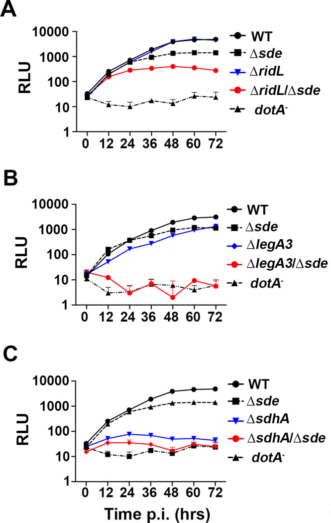
grows intracellularly within a host membrane-bound vacuole that is formed in response to a bacterial type IV secretion system (T4SS). T4SS translocated Sde proteins promote phosphoribosyl-linked ubiquitination of endoplasmic reticulum protein Rtn4, but the role played by this modification is obscure due to lack of clear growth defects of mutants. To identify the steps in vacuole biogenesis promoted by these proteins, mutations were identified that unmasked growth defects in Δ strains. Mutations in the , and genes aggravated the Δ fitness defect, resulting in disruption of the -containing vacuole (LCV) membrane within 2 hrs of bacterial contact with host cells. Depletion of Rab5B and sorting nexin 1 partially bypassed loss of Sde proteins, consistent with Sde blocking early endosome and retrograde trafficking, similar to roles previously demonstrated for SdhA and RidL proteins. Sde protein protection of LCV lysis was only observed shortly after infection, presumably because Sde proteins are inactivated by the metaeffector SidJ during the course of infection. Deletion of SidJ extended the time that Sde proteins could prevent vacuole disruption, indicating that Sde proteins are negatively regulated at the posttranslational level and are limited to protecting membrane integrity at the earliest stages of replication. Transcriptional analysis was consistent with this timing model for an early point of execution of Sde protein. Therefore, Sde proteins act as temporally-regulated vacuole guards during establishment of the replication niche, possibly by constructing a physical barrier that blocks access of disruptive host compartments early during biogenesis of the LCV.
Maintaining replication compartment integrity is critical for growth of intravacuolar pathogens within host cells. By identifying genetically redundant pathways, Sde proteins that promote phosphoribosyl-linked ubiquitination of target eukaryotic proteins are shown to be temporally-regulated vacuole guards, preventing replication vacuole dissolution during early stages of infection. As targeting of reticulon 4 by these proteins leads to tubular endoplasmic reticulum aggregation, Sde proteins are likely to construct a barrier that blocks access of disruptive early endosomal compartments to the replication vacuole. Our study provides a new framework for how vacuole guards function to support biogenesis of the replicative niche.
在宿主膜结合液泡内进行细胞内生长,该液泡是由细菌IV型分泌系统(T4SS)形成的。T4SS转运的Sde蛋白促进内质网蛋白Rtn4的磷酸核糖基连接的泛素化,但由于突变体缺乏明显的生长缺陷,这种修饰所起的作用尚不清楚。为了确定这些蛋白促进液泡生物发生的步骤,鉴定了在Δ菌株中揭示生长缺陷的突变。 、 和 基因中的突变加剧了Δ适应性缺陷,导致在细菌与宿主细胞接触后2小时内含有 的液泡(LCV)膜破裂。Rab5B和分选连接蛋白1的缺失部分绕过了Sde蛋白的缺失,这与Sde阻断早期内体和逆行运输一致,类似于先前对SdhA和RidL蛋白所证明的作用。仅在感染后不久观察到Sde蛋白对LCV裂解的保护作用,推测是因为Sde蛋白在感染过程中被meta效应子SidJ灭活。删除SidJ延长了Sde蛋白防止液泡破坏的时间,表明Sde蛋白在翻译后水平受到负调控,并且仅限于在复制的最早阶段保护膜完整性。转录分析与Sde蛋白执行的早期时间模型一致。因此,Sde蛋白在复制小生境建立期间充当时间调节的液泡保护者,可能是通过构建物理屏障来阻止在LCV生物发生早期破坏性宿主区室的进入。
维持复制区室完整性对于宿主细胞内液泡内病原体的生长至关重要。通过鉴定基因冗余途径,促进靶真核蛋白磷酸核糖基连接泛素化的Sde蛋白被证明是时间调节的液泡保护者,可防止感染早期复制液泡溶解。由于这些蛋白对网质蛋白4的靶向导致管状内质网聚集,Sde蛋白可能构建了一个屏障,阻止破坏性早期内体区室进入复制液泡。我们的研究为液泡保护者如何支持复制小生境的生物发生提供了一个新框架。