• 文献检索
  • 文档翻译
  • 深度研究
  • 学术资讯
  • Suppr Zotero 插件Zotero 插件
  • 邀请有礼
  • 套餐&价格
  • 历史记录
应用&插件
Suppr Zotero 插件Zotero 插件浏览器插件Mac 客户端Windows 客户端微信小程序
定价
高级版会员购买积分包购买API积分包
服务
文献检索文档翻译深度研究API 文档MCP 服务
关于我们
关于 Suppr公司介绍联系我们用户协议隐私条款
关注我们

Suppr 超能文献

核心技术专利:CN118964589B侵权必究
粤ICP备2023148730 号-1Suppr @ 2026

文献检索

告别复杂PubMed语法,用中文像聊天一样搜索,搜遍4000万医学文献。AI智能推荐,让科研检索更轻松。

立即免费搜索

文件翻译

保留排版,准确专业,支持PDF/Word/PPT等文件格式,支持 12+语言互译。

免费翻译文档

深度研究

AI帮你快速写综述,25分钟生成高质量综述,智能提取关键信息,辅助科研写作。

立即免费体验

肝上皮样血管内皮瘤的CT表现及分类

CT appearances and classification of hepatic epithelioid hemangioendothelioma.

作者信息

Tan Haidong, Zhou Ruiquan, Yu Hongwei, Teng Feng, Si Shuang, Liu Liguo, Yang Shiwei, Han Dongdong, Liu Xiaolei

机构信息

Second Department of Hepatopancreatobiliary Surgery, China-Japan Friendship Hospital, 2 Yinghua Dongjie, Hepingli, Beijing, 100029, China.

Department of Radiology, China-Japan Friendship Hospital, Beijing, China.

出版信息

Insights Imaging. 2023 Apr 1;14(1):56. doi: 10.1186/s13244-023-01410-z.

DOI:10.1186/s13244-023-01410-z
PMID:37005950
原文链接:https://pmc.ncbi.nlm.nih.gov/articles/PMC10067783/
Abstract

BACKGROUND

Hepatic epithelioid hemangioendothelioma (HEH) is extremely rare, and CT features have never been analyzed in a large group of patients.

METHODS

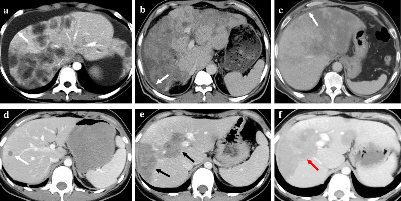
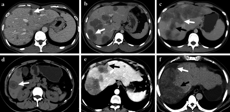
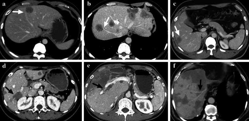
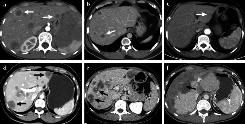
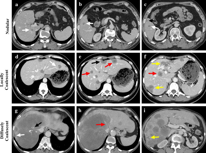
A retrospective study was designed to review the contrast-enhanced CT images of HEH patients. Intrahepatic lesions were categorized into three types: nodular, locally coalescent (coalescent lesion contained in one segment) or diffusely coalescent (coalescent lesion occupied more than one segment). CT features were compared among lesions of different sizes and patients with different lesion types.

RESULTS

A total of 93 HEH patients were included in this study, and 740 lesions were analyzed. The results of per-lesion analysis showed that medium lesions (2-5 cm) had the highest rate of lollipop sign (16.8%) and target-like enhancement (43.1%), while lesions in large group (> 5 cm) had the highest rate of capsular retraction (38.8%) and vascular invasion (38.8%). The differences on enhancement pattern and the rates of lollipop sign and capsular retraction were significant among lesions of different sizes (p < 0.001, respectively). The results of per-patient analysis showed that patients in locally coalescent group had the highest rates of lollipop sign (74.3%) and target sign (94.3%). All patients in diffusely coalescent group had capsular retraction and vascular invasion. CT appearances of capsular retraction, lollipop sign, target sign and vascular invasion differed significantly among patients with different lesion types (p < 0.001, p = 0.005, p = 0.006 and p < 0.001, respectively).

CONCLUSION

CT features variated among HEH patients with different lesion types, and radiological appearances of HEH should be classified into nodular type, locally coalescent type and diffusely coalescent type.

摘要

背景

肝上皮样血管内皮瘤(HEH)极为罕见,且从未在一大组患者中分析过其CT特征。

方法

设计一项回顾性研究,以回顾HEH患者的增强CT图像。肝内病变分为三种类型:结节型、局部融合型(融合病变局限于一个肝段)或弥漫融合型(融合病变占据一个以上肝段)。比较不同大小病变及不同病变类型患者的CT特征。

结果

本研究共纳入93例HEH患者,分析了740个病变。单病变分析结果显示,中等大小病变(2 - 5 cm)的棒棒糖征发生率最高(16.8%)和靶样强化发生率最高(43.1%),而大病变组(>5 cm)的包膜回缩率最高(38.8%)和血管侵犯率最高(38.8%)。不同大小病变的强化方式、棒棒糖征及包膜回缩率差异有统计学意义(p均<0.001)。单患者分析结果显示,局部融合组患者的棒棒糖征发生率最高(74.3%)和靶征发生率最高(94.3%)。弥漫融合组所有患者均有包膜回缩和血管侵犯。不同病变类型患者的包膜回缩、棒棒糖征、靶征及血管侵犯的CT表现差异有统计学意义(p分别为<0.001、=0.005、=0.006和<0.001)。

结论

不同病变类型的HEH患者CT特征存在差异,HEH的影像学表现应分为结节型、局部融合型和弥漫融合型。

https://cdn.ncbi.nlm.nih.gov/pmc/blobs/479f/10067783/7f370bd46ab3/13244_2023_1410_Fig6_HTML.jpg
https://cdn.ncbi.nlm.nih.gov/pmc/blobs/479f/10067783/6431dab26654/13244_2023_1410_Fig1_HTML.jpg
https://cdn.ncbi.nlm.nih.gov/pmc/blobs/479f/10067783/466631b4e05d/13244_2023_1410_Fig2_HTML.jpg
https://cdn.ncbi.nlm.nih.gov/pmc/blobs/479f/10067783/017410bba7d3/13244_2023_1410_Fig3_HTML.jpg
https://cdn.ncbi.nlm.nih.gov/pmc/blobs/479f/10067783/0ed5eb5c46c7/13244_2023_1410_Fig4_HTML.jpg
https://cdn.ncbi.nlm.nih.gov/pmc/blobs/479f/10067783/e6b1ba4a3b8e/13244_2023_1410_Fig5_HTML.jpg
https://cdn.ncbi.nlm.nih.gov/pmc/blobs/479f/10067783/7f370bd46ab3/13244_2023_1410_Fig6_HTML.jpg

相似文献

1
CT appearances and classification of hepatic epithelioid hemangioendothelioma.肝上皮样血管内皮瘤的CT表现及分类
Insights Imaging. 2023 Apr 1;14(1):56. doi: 10.1186/s13244-023-01410-z.
2
MRI appearances of hepatic epithelioid hemangioendothelioma: a retrospective study of 57 patients.肝脏上皮样血管内皮瘤的MRI表现:57例患者的回顾性研究
Insights Imaging. 2022 Apr 5;13(1):65. doi: 10.1186/s13244-022-01213-8.
3
Typical CT and MRI signs of hepatic epithelioid hemangioendothelioma.肝上皮样血管内皮瘤的典型CT和MRI表现。
Oncol Lett. 2016 Mar;11(3):1699-1706. doi: 10.3892/ol.2016.4149. Epub 2016 Jan 26.
4
Spectrum of appearances on CT and MRI of hepatic epithelioid hemangioendothelioma.肝脏上皮样血管内皮瘤的CT和MRI表现谱
BMC Gastroenterol. 2015 Jun 19;15:69. doi: 10.1186/s12876-015-0299-x.
5
CT and MR imaging findings of hepatic epithelioid hemangioendotheliomas: emphasis on single nodular type.肝上皮样血管内皮瘤的CT和MR成像表现:以单结节型为重点
Abdom Imaging. 2015 Mar;40(3):500-9. doi: 10.1007/s00261-014-0229-3.
6
Clinical characteristics and CT features of hepatic epithelioid haemangioendothelioma and comparison with those of liver metastases.肝上皮样血管内皮瘤的临床特征及CT表现与肝转移瘤的比较
Insights Imaging. 2022 Jan 20;13(1):9. doi: 10.1186/s13244-021-01143-x.
7
Hepatic hemangioendothelioma: CT, MR, and FDG-PET-CT in 67 patients-a bi-institutional comprehensive cancer center review.肝血管内皮细胞瘤:67 例患者的 CT、MR 和 FDG-PET-CT——一家综合癌症中心的双机构回顾性研究。
Eur Radiol. 2020 May;30(5):2435-2442. doi: 10.1007/s00330-019-06637-3. Epub 2020 Jan 30.
8
MRI and FDG PET/CT findings of hepatic epithelioid hemangioendothelioma.MRI 和 FDG PET/CT 对肝脏上皮样血管内皮细胞瘤的表现。
Clin Nucl Med. 2013 Feb;38(2):e66-73. doi: 10.1097/RLU.0b013e318266ceca.
9
CT and MRI diagnosis of hepatic epithelioid hemangioendothelioma.CT 和 MRI 诊断肝上皮样血管内皮细胞瘤。
Hepatobiliary Pancreat Dis Int. 2010 Apr;9(2):154-8.
10
MRI of hepatic epithelioid hemangioendothelioma (HEH).肝上皮样血管内皮瘤(HEH)的磁共振成像(MRI)
J Magn Reson Imaging. 2014 Sep;40(3):552-8. doi: 10.1002/jmri.24391. Epub 2013 Nov 13.

引用本文的文献

1
Hepatic epithelioid hemangioendothelioma, a rare liver tumor: a case report and review of the literature.肝上皮样血管内皮瘤,一种罕见的肝脏肿瘤:病例报告及文献综述
Front Oncol. 2025 May 16;15:1522002. doi: 10.3389/fonc.2025.1522002. eCollection 2025.
2
ESR Bridges: imaging and treatment of uncommon liver tumours-a multidisciplinary view.ESR 桥梁:罕见肝脏肿瘤的影像学与治疗——多学科视角
Eur Radiol. 2025 May 9. doi: 10.1007/s00330-025-11662-6.
3
Nab-Paclitaxel Based Chemotherapy in the Treatment of Advanced Epithelioid Hemangioendothelioma: A Single-Institution Experience.

本文引用的文献

1
Sirolimus combined with interferon-alpha 2b therapy for giant hepatic epithelioid hemangioendothelioma: a case report.西罗莫司联合干扰素-α 2b治疗巨大肝脏上皮样血管内皮瘤:一例报告
Front Oncol. 2022 Aug 24;12:972306. doi: 10.3389/fonc.2022.972306. eCollection 2022.
2
Treatment modalities and long-term outcomes of hepatic hemangioendothelioma in the United States.美国肝血管内皮细胞瘤的治疗方式和长期预后。
HPB (Oxford). 2022 Oct;24(10):1688-1696. doi: 10.1016/j.hpb.2022.03.013. Epub 2022 Apr 14.
3
MRI appearances of hepatic epithelioid hemangioendothelioma: a retrospective study of 57 patients.
基于纳米白蛋白结合型紫杉醇的化疗方案治疗晚期上皮样血管内皮瘤:单中心经验
Cancer Manag Res. 2025 Feb 21;17:373-381. doi: 10.2147/CMAR.S508673. eCollection 2025.
4
Long-term prognosis and treatment modalities of hepatic epithelioid hemangioendothelioma: a retrospective study of 228 patients.肝上皮样血管内皮细胞瘤的长期预后和治疗方式:228 例回顾性研究。
BMC Cancer. 2024 Oct 16;24(1):1285. doi: 10.1186/s12885-024-13053-4.
5
I thought it was a hemangioma! A pictorial essay about common and uncommon liver hemangiomas' mimickers.我原以为那是个血管瘤!一篇关于常见及罕见的肝脏血管瘤模仿者的图文并茂的文章。
Insights Imaging. 2024 Sep 19;15(1):228. doi: 10.1186/s13244-024-01745-1.
6
Unusual liver tumors: spectrum of imaging findings with pathologic correlation.罕见肝脏肿瘤:影像表现谱与病理对照
Diagn Interv Radiol. 2025 Mar 3;31(2):58-67. doi: 10.4274/dir.2024.242827. Epub 2024 Jun 10.
7
Short-term outcomes of combined therapy with sirolimus and interferon-alpha 2b for advanced hepatic epithelioid hemangioendothelioma.西罗莫司与α-2b干扰素联合治疗晚期肝脏上皮样血管内皮瘤的短期疗效
Ther Adv Med Oncol. 2024 Jan 3;16:17588359231220509. doi: 10.1177/17588359231220509. eCollection 2024.
8
Cytopathologic features of epithelioid hemangioendothelioma including touch imprints for rapid on-site evaluation.上皮样血管内皮瘤的细胞病理学特征,包括用于快速现场评估的触摸印片。
Cytojournal. 2023 Sep 4;20:29. doi: 10.25259/Cytojournal_57_2022. eCollection 2023.
9
Hepatic epithelioid hemangioendothelioma-a single-institution experience with 51 cases.肝上皮样血管内皮瘤——单机构51例病例经验
Front Oncol. 2023 Aug 3;13:1236134. doi: 10.3389/fonc.2023.1236134. eCollection 2023.
肝脏上皮样血管内皮瘤的MRI表现:57例患者的回顾性研究
Insights Imaging. 2022 Apr 5;13(1):65. doi: 10.1186/s13244-022-01213-8.
4
Novel and Specific MRI Features Indicate the Clinical Features of Patients With Rare Hepatic Tumor Epithelioid Hemangioendothelioma.新颖且独特的MRI特征显示罕见肝肿瘤上皮样血管内皮瘤患者的临床特征。
Front Oncol. 2022 Jan 27;12:729177. doi: 10.3389/fonc.2022.729177. eCollection 2022.
5
Clinical characteristics and CT features of hepatic epithelioid haemangioendothelioma and comparison with those of liver metastases.肝上皮样血管内皮瘤的临床特征及CT表现与肝转移瘤的比较
Insights Imaging. 2022 Jan 20;13(1):9. doi: 10.1186/s13244-021-01143-x.
6
Massive Calcified Epithelioid Hemangioendothelioma With Multifocal Involvement: An Imaging Diagnosis Dilemma and a Rare Case Report.多灶性累及的巨大钙化上皮样血管内皮瘤:影像诊断难题及罕见病例报告
Front Oncol. 2021 Dec 17;11:782970. doi: 10.3389/fonc.2021.782970. eCollection 2021.
7
Efficacy and Safety of Interferon-Alpha 2b for Patients with Hepatic Epithelioid Hemangioendothelioma: Outcomes of a Case-Series Analysis.α-2b干扰素治疗肝上皮样血管内皮瘤患者的疗效与安全性:病例系列分析结果
Cancer Manag Res. 2021 Nov 3;13:8273-8279. doi: 10.2147/CMAR.S334171. eCollection 2021.
8
Liver transplantation for hepatic epithelioid hemangioendothelioma.肝上皮样血管内皮瘤的肝移植
Cancer. 2021 Oct 15;127(20):3714-3716. doi: 10.1002/cncr.33751. Epub 2021 Jul 13.
9
Outcomes of hepatic epithelioid hemangioendothelioma with different managements: a retrospective investigation.不同治疗方式的肝上皮样血管内皮细胞瘤的结局:一项回顾性研究。
Eur Rev Med Pharmacol Sci. 2021 Jun;25(12):4274-4282. doi: 10.26355/eurrev_202106_26133.
10
Epithelioid hemangioendothelioma, an ultra-rare cancer: a consensus paper from the community of experts.上皮样血管内皮细胞瘤,一种超罕见的癌症:来自专家社区的共识文件。
ESMO Open. 2021 Jun;6(3):100170. doi: 10.1016/j.esmoop.2021.100170. Epub 2021 Jun 2.