Huang Jiayan, Gao Ling, Li Jiawu, Yang Rui, Jiang Zhenpeng, Liao Min, Luo Yan, Lu Qiang
Department of Ultrasound, West China Hospital of Sichuan University, Chengdu, China.
Department of Ultrasound, Chengdu BOE Hospital, Chengdu, China.
Front Oncol. 2023 Mar 15;13:1140277. doi: 10.3389/fonc.2023.1140277. eCollection 2023.
To compare the diagnostic efficacy of SonoVue-enhanced and Sonazoid-enhanced ultrasound (US) for hepatocellular carcinoma (HCC) in patients at high risk.
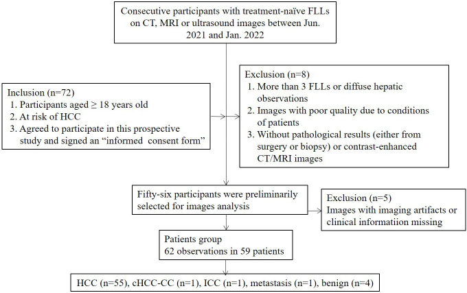
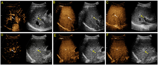
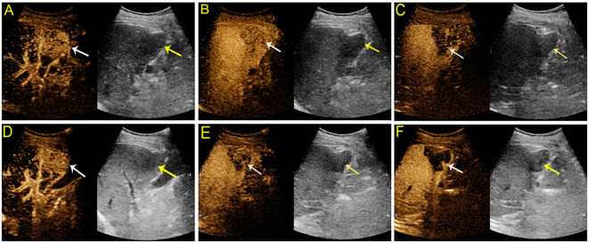
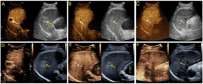
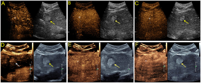
Between August 2021 and February 2022, participants at high risk for HCC with focal liver lesions were enrolled and underwent both SonoVue- and Sonazoid-enhanced US. Vascular-phase and Kupffer phase (KP) imaging features of contrast-enhanced US (CEUS) were analyzed. The diagnostic performance of both contrast agent-enhanced US according to the CEUS liver imaging reporting and data system (LI-RADS) and the modified criteria (using KP defect instead of late and mild washout) were compared. Histopathology and contrast-enhanced MRI/CT were used as reference standards.
In total, 62 nodules, namely, 55 HCCs, 3 non-HCC malignancies and 4 hemangiomas, from 59 participants were included. SonoVue-enhanced US had comparable sensitivity to Sonazoid-enhanced US for diagnosing HCC [80% (95% confidential interval (CI): 67%, 89.6%) versus 74.6% (95% CI: 61%, 85.3%), = 0.25]. Both SonoVue and Sonazoid-enhanced US achieved a specificity of 100%. Compared with CEUS LI-RADS, the modified criteria with Sonazoid did not improve sensitivity for HCC diagnosis [74.6% (95% CI: 61%, 85.3%) versus 76.4% (95% CI: 63%, 86.8%), = 0.99].
Sonazoid-enhanced US had comparable diagnostic performance to SonoVue-enhanced US for patients with HCC risk. KP did not considerably improve the diagnostic efficacy, whereas KP defects in atypical hemangioma may be pitfalls in diagnosing HCC. Further studies with larger sample sizes are needed to further validate the conclusions in the present study.
比较声诺维增强超声和索拉迪德增强超声对高危患者肝细胞癌(HCC)的诊断效能。
2021年8月至2022年2月,纳入有局灶性肝病变的HCC高危参与者,对其进行声诺维和索拉迪德增强超声检查。分析对比增强超声(CEUS)的血管期和库普弗期(KP)成像特征。比较两种造影剂增强超声根据CEUS肝脏成像报告和数据系统(LI-RADS)及改良标准(使用KP缺损代替延迟和轻度廓清)的诊断性能。组织病理学和对比增强MRI/CT用作参考标准。
共纳入59名参与者的62个结节,其中55个HCC、3个非HCC恶性肿瘤和4个血管瘤。声诺维增强超声诊断HCC的敏感性与索拉迪德增强超声相当[80%(95%置信区间(CI):67%,89.6%)对74.6%(95%CI:61%,85.3%),P = 0.25]。声诺维和索拉迪德增强超声的特异性均达到100%。与CEUS LI-RADS相比,索拉迪德的改良标准未提高HCC诊断的敏感性[74.6%(95%CI:61%,85.3%)对76.4%(95%CI:63%,86.8%),P = 0.99]。
对于有HCC风险的患者,索拉迪德增强超声的诊断性能与声诺维增强超声相当。KP未显著提高诊断效能,而非典型血管瘤中的KP缺损可能是诊断HCC的陷阱。需要进一步开展更大样本量的研究以进一步验证本研究结论。