• 文献检索
  • 文档翻译
  • 深度研究
  • 学术资讯
  • Suppr Zotero 插件Zotero 插件
  • 邀请有礼
  • 套餐&价格
  • 历史记录
应用&插件
Suppr Zotero 插件Zotero 插件浏览器插件Mac 客户端Windows 客户端微信小程序
定价
高级版会员购买积分包购买API积分包
服务
文献检索文档翻译深度研究API 文档MCP 服务
关于我们
关于 Suppr公司介绍联系我们用户协议隐私条款
关注我们

Suppr 超能文献

核心技术专利:CN118964589B侵权必究
粤ICP备2023148730 号-1Suppr @ 2026

文献检索

告别复杂PubMed语法,用中文像聊天一样搜索,搜遍4000万医学文献。AI智能推荐,让科研检索更轻松。

立即免费搜索

文件翻译

保留排版,准确专业,支持PDF/Word/PPT等文件格式,支持 12+语言互译。

免费翻译文档

深度研究

AI帮你快速写综述,25分钟生成高质量综述,智能提取关键信息,辅助科研写作。

立即免费体验

改良CEUS LI-RADS在使用Sonazoid诊断肝细胞癌中的应用价值

Usefulness of Modified CEUS LI-RADS for the Diagnosis of Hepatocellular Carcinoma Using Sonazoid.

作者信息

Sugimoto Katsutoshi, Kakegawa Tatsuya, Takahashi Hiroshi, Tomita Yusuke, Abe Masakazu, Yoshimasu Yu, Takeuchi Hirohito, Kasai Yoshitaka, Itoi Takao

机构信息

Department of Gastroenterology and Hepatology, Tokyo Medical University, Tokyo 160-0023, Japan.

出版信息

Diagnostics (Basel). 2020 Oct 15;10(10):828. doi: 10.3390/diagnostics10100828.

DOI:10.3390/diagnostics10100828
PMID:33076435
原文链接:https://pmc.ncbi.nlm.nih.gov/articles/PMC7602639/
Abstract

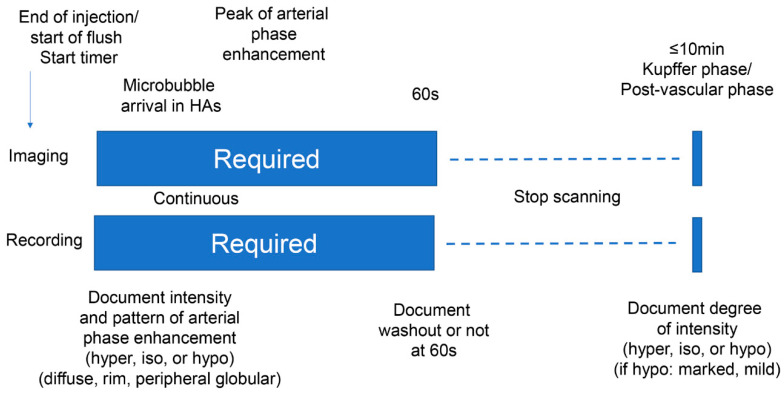
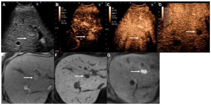
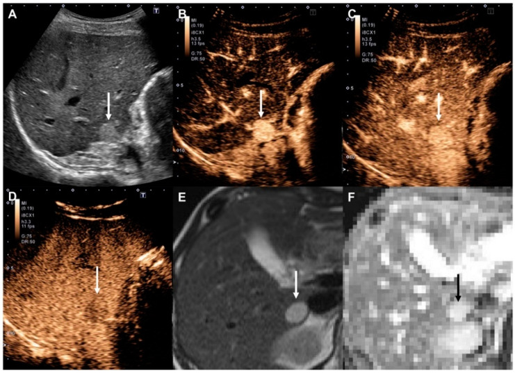
The Contrast-Enhanced Ultrasound Liver Imaging Reporting and Data System (CEUS LI-RADS) was introduced for classifying suspected hepatocellular carcinoma (HCC). However, it cannot be applied to Sonazoid. We assessed the diagnostic usefulness of a modified CEUS LI-RADS for HCC and non-HCC malignancies based on sensitivity, specificity, positive predictive value (PPV), and negative predictive value (NPV). Patients with chronic liver disease at risk for HCC were evaluated retrospectively. Nodules ≥1 cm with arterial phase hyperenhancement, no early washout (within 60 s), and contrast defects in the Kupffer phase were classified as LR-5. Nodules showing early washout, contrast defects in the Kupffer phase, and/or rim enhancement were classified as LR-M. A total of 104 nodules in 104 patients (median age: 70.0 years; interquartile range: 54.5-78.0 years; 74 men) were evaluated. The 48 (46.2%) LR-5 lesions included 45 HCCs, 2 high-flow hemangiomas, and 1 adrenal rest tumor. The PPV of LR-5 for HCC was 93.8% (95% confidence interval (CI): 82.8-98.7%). The 22 (21.2%) LR-M lesions included 16 non-HCC malignancies and 6 HCCs. The PPV of LR-M for non-HCC malignancies, including six intrahepatic cholangiocarcinomas, was 100% (95% CI: 69.8-100%). In conclusion, in the modified CEUS LI-RADS for Sonazoid, LR-5 and LR-M are good predictors of HCC and non-HCC malignancies, respectively.

摘要

对比增强超声肝脏成像报告和数据系统(CEUS LI-RADS)被引入用于对疑似肝细胞癌(HCC)进行分类。然而,它不能应用于Sonazoid。我们基于敏感性、特异性、阳性预测值(PPV)和阴性预测值(NPV)评估了改良的CEUS LI-RADS对HCC和非HCC恶性肿瘤的诊断效用。对有HCC风险的慢性肝病患者进行了回顾性评估。直径≥1 cm且动脉期强化、无早期廓清(60秒内)以及门脉期有对比剂充盈缺损的结节被分类为LR-5。显示早期廓清、门脉期对比剂充盈缺损和/或边缘强化的结节被分类为LR-M。共评估了104例患者的104个结节(中位年龄:70.0岁;四分位间距:54.5 - 78.0岁;男性74例)。48个(46.2%)LR-5病变包括45例HCC、2例高流量血管瘤和1例肾上腺残余肿瘤。LR-5对HCC的PPV为93.8%(95%置信区间(CI):82.8 - 98.7%)。22个(21.2%)LR-M病变包括16例非HCC恶性肿瘤和6例HCC。LR-M对包括6例肝内胆管癌在内的非HCC恶性肿瘤的PPV为100%(95% CI:69.8 - 100%)。总之,在用于Sonazoid的改良CEUS LI-RADS中,LR-5和LR-M分别是HCC和非HCC恶性肿瘤的良好预测指标。

https://cdn.ncbi.nlm.nih.gov/pmc/blobs/6722/7602639/d6c3c35acad3/diagnostics-10-00828-g004.jpg
https://cdn.ncbi.nlm.nih.gov/pmc/blobs/6722/7602639/e041267c999f/diagnostics-10-00828-g001.jpg
https://cdn.ncbi.nlm.nih.gov/pmc/blobs/6722/7602639/595c72eb2923/diagnostics-10-00828-g002.jpg
https://cdn.ncbi.nlm.nih.gov/pmc/blobs/6722/7602639/d22231d6ea16/diagnostics-10-00828-g003.jpg
https://cdn.ncbi.nlm.nih.gov/pmc/blobs/6722/7602639/d6c3c35acad3/diagnostics-10-00828-g004.jpg

相似文献

1
Usefulness of Modified CEUS LI-RADS for the Diagnosis of Hepatocellular Carcinoma Using Sonazoid.改良CEUS LI-RADS在使用Sonazoid诊断肝细胞癌中的应用价值
Diagnostics (Basel). 2020 Oct 15;10(10):828. doi: 10.3390/diagnostics10100828.
2
The efficacy of modified contrast-enhanced ultrasound Liver Imaging Reporting and Data System (CEUS LI-RADS) using Sonazoid in diagnosis of hepatocellular carcinoma: a systematic review and meta-analysis.使用声诺维的改良对比增强超声肝脏影像报告和数据系统(CEUS LI-RADS)在肝细胞癌诊断中的疗效:一项系统评价和荟萃分析。
Quant Imaging Med Surg. 2024 Apr 3;14(4):2927-2937. doi: 10.21037/qims-23-1184. Epub 2024 Mar 21.
3
Sonazoid-enhanced ultrasonography: comparison with CT/MRI Liver Imaging Reporting and Data System in patients with suspected hepatocellular carcinoma.声诺维增强超声检查:与疑似肝细胞癌患者的CT/MRI肝脏影像报告和数据系统的比较
Ultrasonography. 2021 Oct;40(4):486-498. doi: 10.14366/usg.20120. Epub 2021 Jan 15.
4
Comparison of modified CEUS LI-RADS with sonazoid and CT/MRI LI-RADS for diagnosis of hepatocellular carcinoma.改良的超声造影肝脏影像报告和数据系统(CEUS LI-RADS)与声诺维及CT/MRI肝脏影像报告和数据系统(LI-RADS)在肝细胞癌诊断中的比较
Hepatol Res. 2022 Aug;52(8):730-738. doi: 10.1111/hepr.13793. Epub 2022 May 28.
5
Diagnostic accuracy of contrast-enhanced ultrasound for the differential diagnosis of hepatocellular carcinoma: ESCULAP versus CEUS-LI-RADS.超声造影对肝细胞癌鉴别诊断的诊断准确性:爱克发与CEUS-LI-RADS对比
Eur J Gastroenterol Hepatol. 2017 Sep;29(9):1036-1044. doi: 10.1097/MEG.0000000000000916.
6
Contrast ultrasound LI-RADS LR-5 identifies hepatocellular carcinoma in cirrhosis in a multicenter restropective study of 1,006 nodules.在一项多中心回顾性研究中,1006 个结节的对比超声 LI-RADS LR-5 可识别肝硬化中的肝细胞癌。
J Hepatol. 2018 Mar;68(3):485-492. doi: 10.1016/j.jhep.2017.11.007. Epub 2017 Nov 11.
7
Can contrast enhanced ultrasound differentiate intrahepatic cholangiocarcinoma from hepatocellular carcinoma?增强超声能区分肝内胆管细胞癌和肝细胞癌吗?
World J Gastroenterol. 2020 Jul 21;26(27):3938-3951. doi: 10.3748/wjg.v26.i27.3938.
8
Diagnostic Accuracy of CEUS LI-RADS for the Characterization of Liver Nodules 20 mm or Smaller in Patients at Risk for Hepatocellular Carcinoma.CEUS LI-RADS 在肝癌风险患者中对 20mm 或更小的肝脏结节的特征诊断准确性。
Radiology. 2020 Feb;294(2):329-339. doi: 10.1148/radiol.2019191086. Epub 2019 Dec 3.
9
Contrast-enhanced ultrasound Liver Imaging Reporting and Data System: Lights and shadows in hepatocellular carcinoma and cholangiocellular carcinoma diagnosis.超声造影肝脏影像报告与数据系统:在肝细胞癌和胆管细胞癌诊断中的优缺点。
World J Gastroenterol. 2022 Jul 21;28(27):3488-3502. doi: 10.3748/wjg.v28.i27.3488.
10
Contrast-Enhanced Ultrasound Using Perfluorobutane: Impact of Proposed Modified LI-RADS Criteria on Hepatocellular Carcinoma Detection.使用全氟丁烷的超声造影:拟议改良 LI-RADS 标准对肝细胞癌检测的影响。
AJR Am J Roentgenol. 2022 Sep;219(3):434-443. doi: 10.2214/AJR.22.27521. Epub 2022 Apr 20.

引用本文的文献

1
Contrast-Enhanced Sonography of the Liver: How to Avoid Artifacts.肝脏超声造影:如何避免伪像
Diagnostics (Basel). 2024 Aug 20;14(16):1817. doi: 10.3390/diagnostics14161817.
2
The American College of Radiology contrast-enhanced ultrasound Liver Imaging Reporting and Data System and its modified version in diagnosing hepatocellular carcinoma via Sonazoid: a meta-analysis.美国放射学会对比增强超声肝脏影像报告和数据系统及其改良版本在通过声诺维诊断肝细胞癌中的应用:一项荟萃分析。
Quant Imaging Med Surg. 2024 Jul 1;14(7):4555-4566. doi: 10.21037/qims-23-1459. Epub 2024 Jun 27.
3
Compared with SonoVue LR-5, Sonazoid modified LR-5 has better diagnostic sensitivity for hepatocellular carcinoma: a systematic review and meta-analysis.

本文引用的文献

1
Diagnostic Value of Imaging Methods in the Histological Four Grading of Hepatocellular Carcinoma.影像学方法在肝细胞癌组织学四级分级中的诊断价值
Diagnostics (Basel). 2020 May 19;10(5):321. doi: 10.3390/diagnostics10050321.
2
The Impact of Tumor Differentiation on the Prognosis of HBV-Associated Solitary Hepatocellular Carcinoma Following Hepatectomy: A Propensity Score Matching Analysis.肿瘤分化对肝癌切除术治疗后 HBV 相关单发肝癌预后的影响:倾向评分匹配分析。
Dig Dis Sci. 2018 Jul;63(7):1962-1969. doi: 10.1007/s10620-018-5077-5. Epub 2018 May 7.
3
Contrast Enhanced Ultrasound (CEUS) Liver Imaging Reporting and Data System (LI-RADS®): the official version by the American College of Radiology (ACR).
与声诺维LR-5相比,Sonazoid改良的LR-5对肝细胞癌具有更好的诊断敏感性:一项系统评价和荟萃分析。
Quant Imaging Med Surg. 2024 Apr 3;14(4):2978-2992. doi: 10.21037/qims-23-1616. Epub 2024 Mar 13.
4
The efficacy of modified contrast-enhanced ultrasound Liver Imaging Reporting and Data System (CEUS LI-RADS) using Sonazoid in diagnosis of hepatocellular carcinoma: a systematic review and meta-analysis.使用声诺维的改良对比增强超声肝脏影像报告和数据系统(CEUS LI-RADS)在肝细胞癌诊断中的疗效:一项系统评价和荟萃分析。
Quant Imaging Med Surg. 2024 Apr 3;14(4):2927-2937. doi: 10.21037/qims-23-1184. Epub 2024 Mar 21.
5
Intra-individual comparison of Sonazoid contrast-enhanced ultrasound and SonoVue contrast-enhanced ultrasound in diagnosing hepatocellular carcinoma.超声造影剂声诺维与声诺美增强超声在诊断肝细胞癌中的个体内比较。
Abdom Radiol (NY). 2024 May;49(5):1432-1443. doi: 10.1007/s00261-024-04250-7. Epub 2024 Apr 7.
6
Diagnosis of hepatocellular carcinoma using Sonazoid: a comprehensive review.使用Sonazoid诊断肝细胞癌:全面综述
J Liver Cancer. 2023 Sep;23(2):272-283. doi: 10.17998/jlc.2023.08.25. Epub 2023 Sep 19.
7
Sonazoid-enhanced ultrasonography for noninvasive imaging diagnosis of hepatocellular carcinoma: special emphasis on the 2022 KLCA-NCC guideline.用于肝细胞癌无创成像诊断的声诺维增强超声检查:特别强调2022年KLCA-NCC指南
Ultrasonography. 2023 Oct;42(4):479-489. doi: 10.14366/usg.23051. Epub 2023 Jun 15.
8
Modified CEUS LI-RADS using Sonazoid for the diagnosis of hepatocellular carcinoma.使用声诺维的改良CEUS LI-RADS用于肝细胞癌的诊断。
Ultrasonography. 2023 Jul;42(3):388-399. doi: 10.14366/usg.23065. Epub 2023 May 31.
9
Diagnosing Hepatocellular Carcinoma Using Sonazoid Contrast-Enhanced Ultrasonography: 2023 Guidelines From the Korean Society of Radiology and the Korean Society of Abdominal Radiology.使用 SonoVue 对比增强超声诊断肝细胞癌:韩国放射学会和韩国腹部放射学会 2023 年指南。
Korean J Radiol. 2023 Jun;24(6):482-497. doi: 10.3348/kjr.2023.0324.
10
Contrast-enhanced ultrasound for the diagnosis of hepatocellular carcinoma in adults with chronic liver disease.对比增强超声在慢性肝病成人肝细胞癌诊断中的应用。
Cochrane Database Syst Rev. 2022 Sep 2;9(9):CD013483. doi: 10.1002/14651858.CD013483.pub2.
对比增强超声(CEUS)肝脏影像报告和数据系统(LI-RADS®):美国放射学会(ACR)的官方版本。
Ultraschall Med. 2017 Jan;38(1):85-86. doi: 10.1055/s-0042-124369. Epub 2017 Mar 1.
4
High-flow haemangiomas versus hypervascular hepatocellular carcinoma showing "pseudo-washout" on gadoxetic acid-enhanced hepatic MRI: value of diffusion-weighted imaging in the differential diagnosis of small lesions.钆塞酸增强肝脏磁共振成像上表现为“假性廓清”的高流量血管瘤与高血供肝细胞癌:扩散加权成像在小病灶鉴别诊断中的价值
Clin Radiol. 2017 Mar;72(3):247-254. doi: 10.1016/j.crad.2016.09.020. Epub 2016 Oct 24.
5
Ultrasound diagnostic criteria for hepatic tumors.肝脏肿瘤的超声诊断标准。
J Med Ultrason (2001). 2014 Jan;41(1):113-23. doi: 10.1007/s10396-013-0500-1.
6
Contrast-Enhanced Ultrasound for the Characterization of Hepatocellular Carcinoma and Intrahepatic Cholangiocarcinoma.对比增强超声用于肝细胞癌和肝内胆管癌的特征性诊断
Liver Cancer. 2015 Dec;4(4):241-52. doi: 10.1159/000367738. Epub 2015 Oct 21.
7
Kupffer-phase findings of hepatic hemangiomas in contrast-enhanced ultrasound with sonazoid.使用声诺维增强超声检查肝血管瘤的库普弗期表现。
Ultrasound Med Biol. 2014 Jun;40(6):1089-95. doi: 10.1016/j.ultrasmedbio.2013.12.019. Epub 2014 Feb 17.
8
Position paper of the Italian Association for the Study of the Liver (AISF): the multidisciplinary clinical approach to hepatocellular carcinoma.意大利肝脏研究协会(AISF)立场文件:肝细胞癌的多学科临床方法。
Dig Liver Dis. 2013 Sep;45(9):712-23. doi: 10.1016/j.dld.2013.01.012. Epub 2013 Feb 23.
9
Guidelines and good clinical practice recommendations for Contrast Enhanced Ultrasound (CEUS) in the liver - update 2012: A WFUMB-EFSUMB initiative in cooperation with representatives of AFSUMB, AIUM, ASUM, FLAUS and ICUS.肝脏对比增强超声(CEUS)指南和临床实践良好建议-2012 年更新:WFUMB-EFSUMB 与 AFSUMB、AIUM、ASUM、FLAUS 和 ICUS 的代表合作开展的一项倡议。
Ultrasound Med Biol. 2013 Feb;39(2):187-210. doi: 10.1016/j.ultrasmedbio.2012.09.002. Epub 2012 Nov 5.
10
Guidelines and good clinical practice recommendations for contrast enhanced ultrasound (CEUS) in the liver--update 2012: a WFUMB-EFSUMB initiative in cooperation with representatives of AFSUMB, AIUM, ASUM, FLAUS and ICUS.肝脏对比增强超声(CEUS)指南和临床实践良好建议-2012 年更新:WFUMB-EFSUMB 与 AFSUMB、AIUM、ASUM、FLAUS 和 ICUS 的代表合作开展的一项倡议。
Ultraschall Med. 2013 Feb;34(1):11-29. doi: 10.1055/s-0032-1325499. Epub 2012 Nov 5.