TU Dresden, Department of Medical Physics and Biomedical Engineering, Faculty of Medicine, Dresden, Germany.
TU Dresden, Anesthesiology and Intensive Care Medicine, Clinical Sensoring and Monitoring, Faculty of Medicine, Dresden, Germany.
J Biomed Opt. 2023 Dec;28(12):121203. doi: 10.1117/1.JBO.28.12.121203. Epub 2023 Mar 29.
Endoscopic optical coherence tomography (OCT) is of growing interest for diagnostics of the tympanic membrane (TM) and the middle ear but generally lacks a tissue-specific contrast.
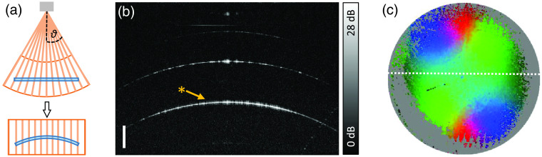
To assess the collagen fiber layer within the TM, an endoscopic imaging method utilizing the polarization changes induced by the birefringent connective tissue was developed.
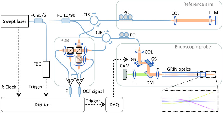
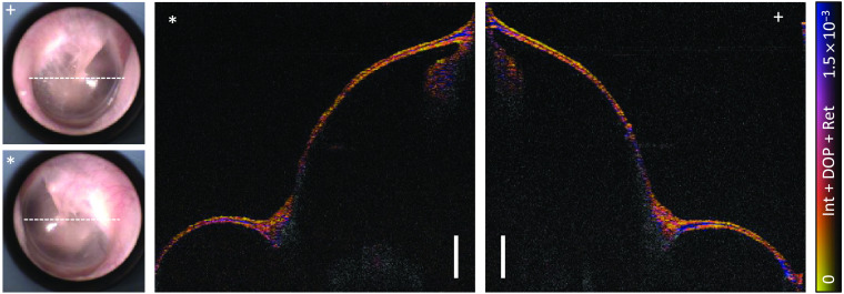
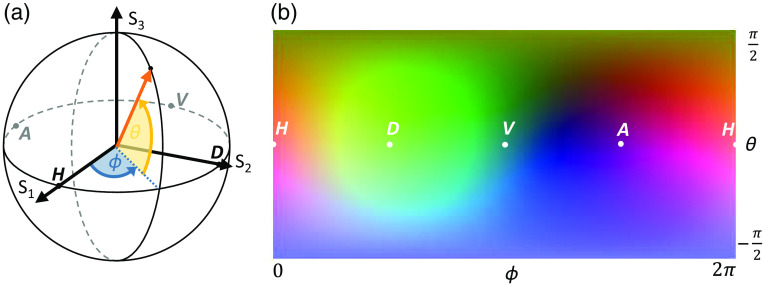
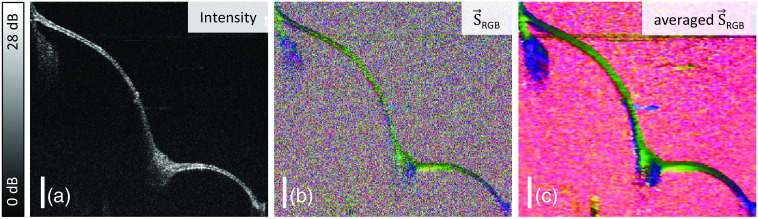
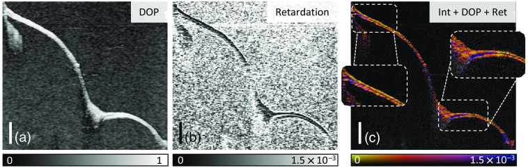
An endoscopic swept-source OCT setup was redesigned and extended by a polarization-diverse balanced detection unit. Polarization-sensitive OCT (PS-OCT) data were visualized by a differential Stokes-based processing and the derived local retardation. The left and right ears of a healthy volunteer were examined.
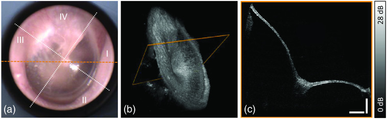
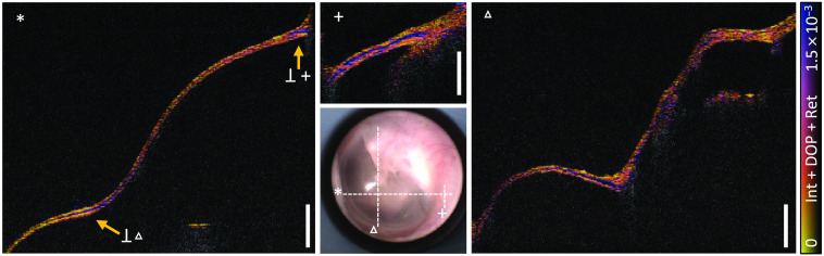
Distinct retardation signals in the annulus region of the TM and near the umbo revealed the layered structure of the TM. Due to the TM's conical shape and orientation in the ear canal, high incident angles onto the TM's surface, and low thicknesses compared to the axial resolution limit of the system, other regions of the TM were more difficult to evaluate.
The use of endoscopic PS-OCT is feasible to differentiate birefringent and nonbirefringent tissue of the human TM . Further investigations on healthy as well as pathologically altered TMs are required to validate the diagnostic potential of this technique.
内窥镜光学相干断层扫描(OCT)对于诊断鼓膜(TM)和中耳越来越感兴趣,但通常缺乏组织特异性对比。
为了评估 TM 内的胶原纤维层,开发了一种利用双折射结缔组织引起的偏振变化的内窥镜成像方法。
重新设计了内窥镜扫频源 OCT 装置,并通过偏振变化的平衡检测单元进行了扩展。偏振敏感 OCT(PS-OCT)数据通过基于差分斯托克斯的处理和推导的局部延迟进行可视化。检查了一名健康志愿者的左耳和右耳。
TM 环状区和耳屏附近的明显延迟信号揭示了 TM 的分层结构。由于 TM 的锥形形状和在耳道中的方向、高入射角到 TM 表面以及与系统轴向分辨率限制相比的低厚度,TM 的其他区域更难以评估。
使用内窥镜 PS-OCT 可以区分人类 TM 的双折射和非双折射组织。需要对健康以及病理改变的 TM 进行进一步研究,以验证该技术的诊断潜力。