Niu Xiaoyu, Gao Xinyu, Lv Qingqing, Zhang Mengzhe, Dang Jinghan, Sun Jieping, Wang Weijian, Wei Yarui, Cheng Jingliang, Han Shaoqiang, Zhang Yong
Department of Magnetic Resonance Imaging, The First Affiliated Hospital of Zhengzhou University, Zhengzhou, Henan, China.
Key Laboratory of Magnetic Resonance and Brain Function of Henan Province, Zhengzhou, China.
Front Hum Neurosci. 2023 Mar 17;17:1153976. doi: 10.3389/fnhum.2023.1153976. eCollection 2023.
Chronic smokers have abnormal spontaneous regional activity and disrupted functional connectivity as revealed by previous neuroimaging studies. Combining different dimensions of resting-state functional indicators may help us learn more about the neuropathological mechanisms of smoking.
The amplitude of low frequency fluctuations (ALFF) of 86 male smokers and 56 male non-smokers were first calculated. Brain regions that displayed significant differences in ALFF between two groups were selected as seeds for further functional connectivity analysis. Besides, we examined correlations between brain areas with abnormal activity and smoking measurements.
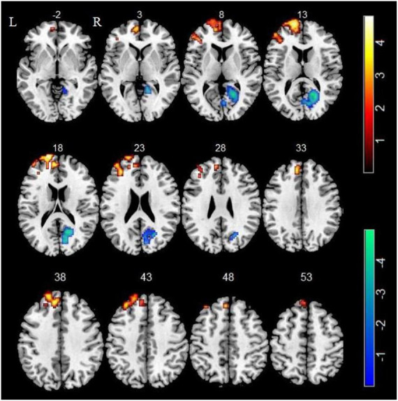
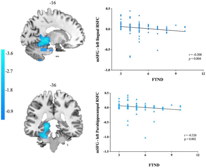
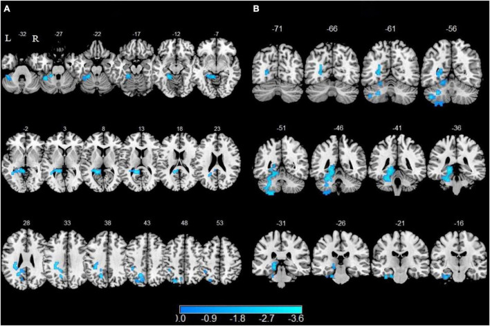
Increased ALFF in left superior frontal gyrus (SFG), left medial superior frontal gyrus (mSFG) and middle frontal gyrus (MFG) as well as decreased ALFF in right calcarine sulcus were observed in smokers compared with non-smokers. In the seed-based functional connectivity analysis, smokers showed attenuated functional connectivity with left SFG in left precuneus, left fusiform gyrus, left lingual gyrus, left cerebellum 4 5 and cerebellum 6 as well as lower functional connectivity with left mSGF in left fusiform gyrus, left lingual gyrus, left parahippocampal gyrus (PHG), left calcarine sulcus, left cerebellum 4 5, cerebellum 6 and cerebellum 8 (GRF corrected, Pvoxel < 0.005, Pcluster<0.05). Furthermore, attenuated functional connectivity with left mSGF in left lingual gyrus and PHG displayed a negative correlation with FTND scores ( = -0.308, = 0.004; = -0.326, = 0.002 Bonferroni corrected).
Our findings of increased ALFF in SFG with reduced functional connectivity to visual attention areas and cerebellum subregions may shed new light on the pathophysiology of smoking.
既往神经影像学研究显示,慢性吸烟者存在异常的自发局部活动和功能连接中断。结合静息态功能指标的不同维度可能有助于我们更深入地了解吸烟的神经病理机制。
首先计算86名男性吸烟者和56名男性非吸烟者的低频振幅(ALFF)。选择两组间ALFF存在显著差异的脑区作为种子点进行进一步的功能连接分析。此外,我们还研究了活动异常脑区与吸烟指标之间的相关性。
与非吸烟者相比,吸烟者左侧额上回(SFG)、左侧额内侧回(mSFG)和额中回(MFG)的ALFF增加,右侧距状沟的ALFF降低。在基于种子点的功能连接分析中,吸烟者在左侧楔前叶、左侧梭状回、左侧舌回、左侧小脑4、5区和小脑6区与左侧SFG的功能连接减弱,在左侧梭状回、左侧舌回、左侧海马旁回(PHG)、左侧距状沟、左侧小脑4、5区、小脑6区和小脑8区与左侧mSGF的功能连接降低(GRF校正,P体素<0.005,P簇<0.05)。此外,左侧舌回和PHG与左侧mSGF的功能连接减弱与FTND评分呈负相关(=-0.308,=0.004;=-0.326,=0.002,Bonferroni校正)。
我们发现SFG的ALFF增加,与视觉注意力区域和小脑亚区的功能连接减少,这可能为吸烟的病理生理学提供新的线索。