Huang Xin, Cai Feng-Qin, Hu Pei-Hong, Zhong Yu-Lin, Zhang Ying, Wei Rong, Pei Chong-Gang, Zhou Fu-Qing, Shao Yi
Department of Ophthalmology, Jiangxi Province Clinical Ophthalmology Institute, First Affiliated Hospital of Nanchang University, Nanchang, People's Republic of China ; Department of Ophthalmology, First People's Hospital of Jiujiang, Jiujiang, People's Republic of China.
Department of Radiology, First Affiliated Hospital of Nanchang University, Nanchang, People's Republic of China.
Neuropsychiatr Dis Treat. 2015 Dec 16;11:3075-83. doi: 10.2147/NDT.S92497. eCollection 2015.
To use the amplitude of low-frequency fluctuation (ALFF) technique to investigate the local features of spontaneous brain activity in optic neuritis (ON) and their relationship with behavioral performance.
Twelve patients with ON (four male, eight female) and twelve age-, sex-, and education status-matched healthy controls (HCs) (four male, eight female) underwent resting-state functional magnetic resonance imaging (rs-fMRI) scans. The ALFF technique was used to assess local features of spontaneous brain activity. Correlation analysis was used to explore the relationship between the observed mean ALFF values of the different areas and visual evoked potentials (VEPs) in patients with ON.
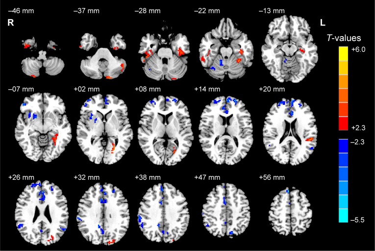
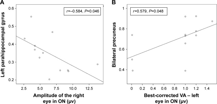
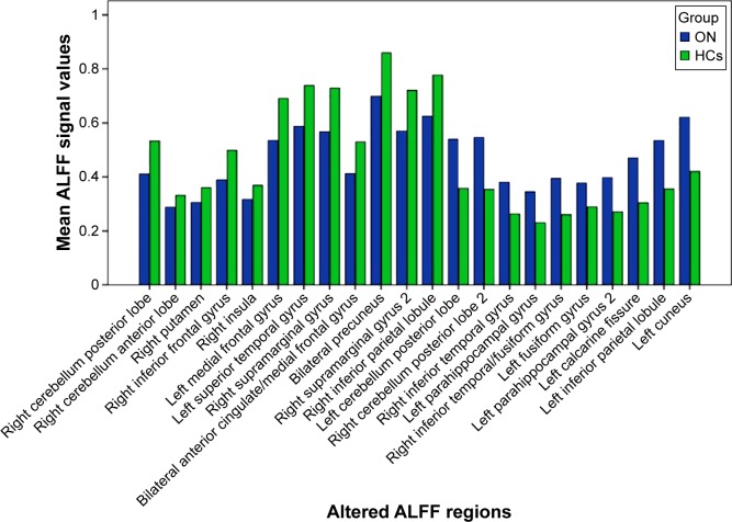
Compared with HCs, patients with ON had significantly decreased ALFF values in the posterior and anterior lobes of the right cerebellum, right putamen, right inferior frontal gyrus, right insula, right supramarginal gyrus, right inferior parietal lobule, left medial frontal gyrus, left superior temporal gyrus, bilateral anterior cingulate/medial frontal gyrus, and bilateral precuneus, and significantly increased ALFF values in the posterior lobes of the left and right cerebellum, right inferior temporal gyrus, right inferior temporal/fusiform gyrus, left parahippocampal gyrus, left fusiform gyrus, left calcarine fissure, left inferior parietal lobule, and left cuneus. We found negative correlations between the mean ALFF signal value of the left parahippocampal gyrus and the VEP amplitude of the right eye in ON (r=-0.584, P=0.046), and a positive correlation between the mean ALFF signal value of the bilateral precuneus and the best-corrected visual acuity of the left eye (r=0.579, P=0.048) in patients with ON.
ON mainly seems to involve dysfunction in the default-mode network, cerebellum, and limbic system, which may reflect the underlying pathologic mechanism of ON.
运用低频振幅(ALFF)技术研究视神经炎(ON)患者脑自发活动的局部特征及其与行为表现的关系。
12例ON患者(4例男性,8例女性)和12名年龄、性别及教育程度相匹配的健康对照者(HCs)(4例男性,8例女性)接受静息态功能磁共振成像(rs-fMRI)扫描。采用ALFF技术评估脑自发活动的局部特征。相关性分析用于探讨ON患者不同脑区观察到的平均ALFF值与视觉诱发电位(VEP)之间的关系。
与HCs相比,ON患者右侧小脑后叶和前叶、右侧壳核、右侧额下回、右侧脑岛、右侧缘上回、右侧顶下小叶、左侧额内侧回、左侧颞上回、双侧前扣带回/额内侧回以及双侧楔前叶的ALFF值显著降低,而左侧和右侧小脑后叶、右侧颞下回、右侧颞下回/梭状回、左侧海马旁回、左侧梭状回、左侧距状裂、左侧顶下小叶以及左侧楔叶的ALFF值显著升高。我们发现ON患者左侧海马旁回的平均ALFF信号值与右眼VEP振幅呈负相关(r = -0.584,P = 0.046),双侧楔前叶的平均ALFF信号值与左眼最佳矫正视力呈正相关(r = 0.579,P = 0.048)。
ON似乎主要涉及默认模式网络、小脑和边缘系统功能障碍,这可能反映了ON潜在的病理机制。