Division of Neuroradiology, Department of Radiology, University of Michigan, 1500 E Medical Center Dr, Ann Arbor, MI, 48109, USA.
Department of Radiology, Graduate School of Medicine, The University of Tokyo, 7-3-1, Hongo, Bunkyo-ku, Tokyo, 113-8655, Japan.
Jpn J Radiol. 2023 Sep;41(9):911-927. doi: 10.1007/s11604-023-01417-y. Epub 2023 Apr 3.
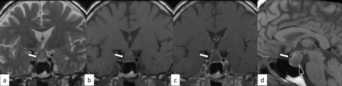
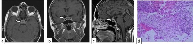
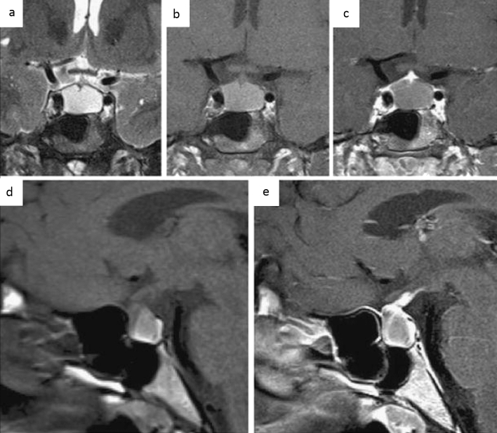
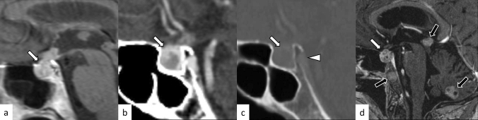
Hypophysitis is an inflammatory disease affecting the pituitary gland. Hypophysitis can be classified into multiple types depending on the mechanisms (primary or secondary), histology (lymphocytic, granulomatous, xanthomatous, plasmacytic/IgG4 related, necrotizing, or mixed), and anatomy (adenohypophysitis, infundibulo-neurohypophysitis, or panhypophysitis). An appropriate diagnosis is vital for managing these potentially life-threatening conditions. However, physiological morphological alterations, remnants, and neoplastic and non-neoplastic lesions may masquerade as hypophysitis, both clinically and radiologically. Neuroimaging, as well as imaging findings of other sites of the body, plays a pivotal role in diagnosis. In this article, we will review the types of hypophysitis and summarize clinical and imaging features of both hypophysitis and its mimickers.
垂体炎是一种影响垂体的炎症性疾病。根据发病机制(原发性或继发性)、组织学(淋巴细胞性、肉芽肿性、含铁血黄素沉积性、浆细胞/IgG4 相关性、坏死性或混合性)和解剖学(腺垂体炎、漏斗神经垂体炎或全垂体炎),垂体炎可分为多种类型。适当的诊断对于治疗这些潜在危及生命的疾病至关重要。然而,生理形态改变、残留物以及肿瘤性和非肿瘤性病变可能在临床上和影像学上表现为垂体炎。神经影像学以及身体其他部位的影像学表现对诊断起着关键作用。本文将回顾垂体炎的类型,并总结垂体炎及其类似物的临床和影像学特征。