Department of Neurology, University Hospital Essen, Hufelandstrasse 55, 45147, Essen, Germany.
Center for Translational Neuro- and Behavioral Scienes (C-TNBS), University Hospital Essen, Essen, Germany.
J Neurol. 2023 Jul;270(7):3483-3491. doi: 10.1007/s00415-023-11689-z. Epub 2023 Apr 4.
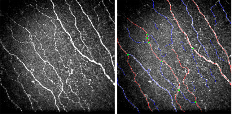
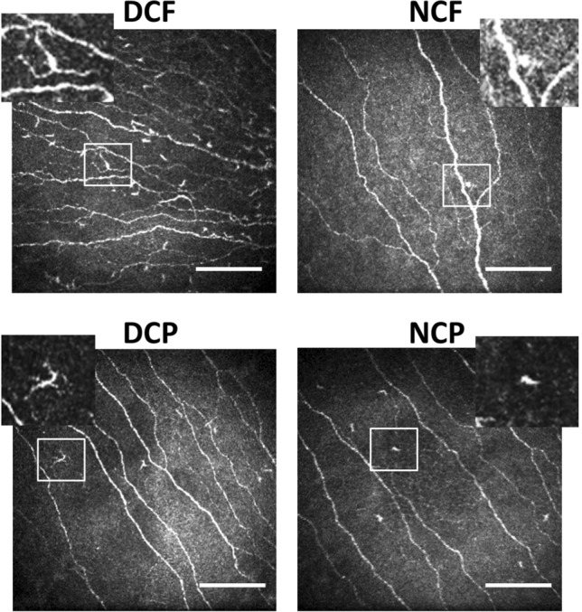
Hereditary transthyretin amyloidosis (ATTRv amyloidosis) is a rare, but life-threatening protein misfolding disorder due to TTR gene mutations. Cardiomyopathy (ATTRv-CM) and polyneuropathy (ATTRv-PN) with early small nerve fibre involvement are the most common manifestations. Timely diagnosis and treatment initiation are key to limiting progression of disease. Corneal confocal microscopy (CCM) is a non-invasive method to quantify corneal small nerve fibres and immune cell infiltrates in vivo.
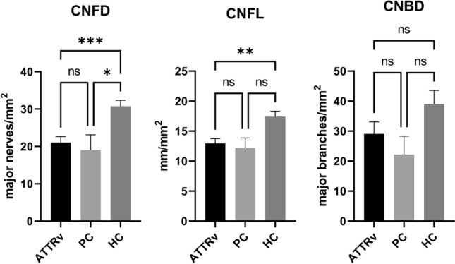
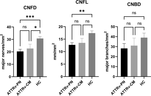
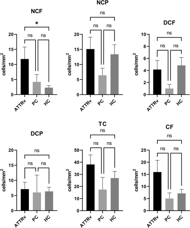
This cross-sectional study investigated the utility of CCM in 20 patients with ATTRv amyloidosis (ATTRv-CM, n = 6; ATTRv-PN, n = 14) and presymptomatic carriers (n = 5) compared to 20 age- and sex-matched healthy controls. Corneal nerve fibre density, corneal nerve fibre length, corneal nerve branch density, and cell infiltrates were assessed.
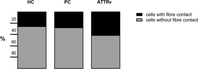
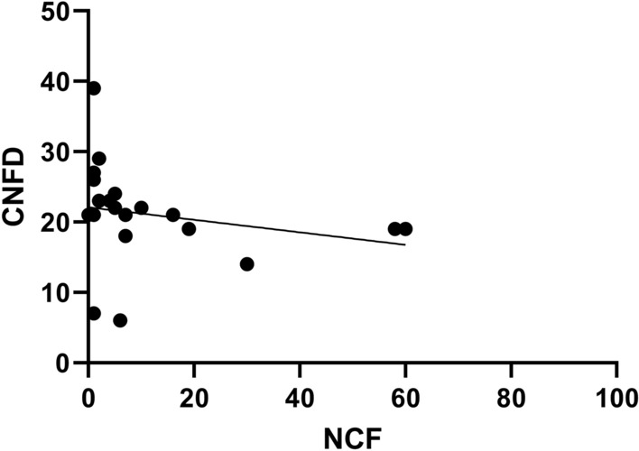
Corneal nerve fibre density and nerve fibre length were significantly lower in patients with ATTRv amyloidosis compared to healthy controls regardless of the clinical phenotype (ATTRv-CM, ATTRv-PN) and corneal nerve fibre density was significantly lower in presymptomatic carriers. Immune cell infiltrates were only evident in patients with ATTRv amyloidosis, which correlated with reduced corneal nerve fibre density.
CCM identifies small nerve fibre damage in presymptomatic carriers and symptomatic patients with ATTRv amyloidosis and may serve as a predictive surrogate marker to identify individuals at risk of developing symptomatic amyloidosis. Furthermore, increased corneal cell infiltration suggests an immune-mediated mechanism in the pathogenesis of amyloid neuropathy.
遗传性转甲状腺素蛋白淀粉样变性(ATTRv 淀粉样变性)是一种罕见但危及生命的蛋白质错误折叠疾病,由 TTR 基因突变引起。心肌病(ATTRv-CM)和多发性神经病(ATTRv-PN)伴早期小纤维神经受累是最常见的表现。及时诊断和开始治疗是限制疾病进展的关键。角膜共焦显微镜(CCM)是一种非侵入性方法,可定量测量活体角膜小神经纤维和免疫细胞浸润。
本横断面研究调查了 CCM 在 20 例 ATTRv 淀粉样变性患者(ATTRv-CM,n=6;ATTRv-PN,n=14)和无症状携带者(n=5)中的效用,并与 20 名年龄和性别匹配的健康对照进行比较。评估了角膜神经纤维密度、角膜神经纤维长度、角膜神经分支密度和细胞浸润。
无论临床表型(ATTRv-CM、ATTRv-PN)如何,ATTRv 淀粉样变性患者的角膜神经纤维密度和神经纤维长度均明显低于健康对照组,而无症状携带者的角膜神经纤维密度明显降低。免疫细胞浸润仅在 ATTRv 淀粉样变性患者中可见,与角膜神经纤维密度降低相关。
CCM 可识别无症状携带者和有症状的 ATTRv 淀粉样变性患者的小神经纤维损伤,可作为预测淀粉样变性风险的替代标志物。此外,角膜细胞浸润增加提示淀粉样神经病变发病机制中存在免疫介导的机制。