Department of Orthopedics, Shanghai Changhai Hospital, Naval Medical University, Shanghai 200433, China.
Institute of Translational Medicine, Shanghai University, Shanghai 200444, China.
Sci Adv. 2023 Apr 5;9(14):eabo7868. doi: 10.1126/sciadv.abo7868.
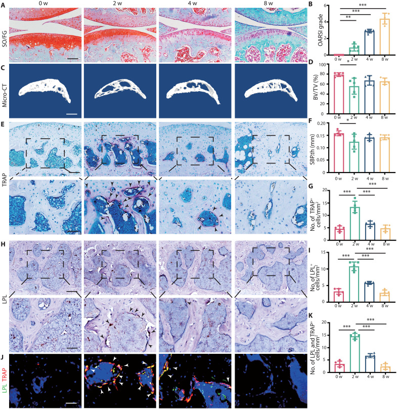
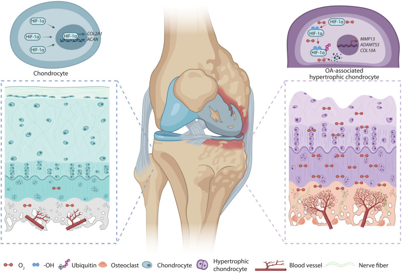
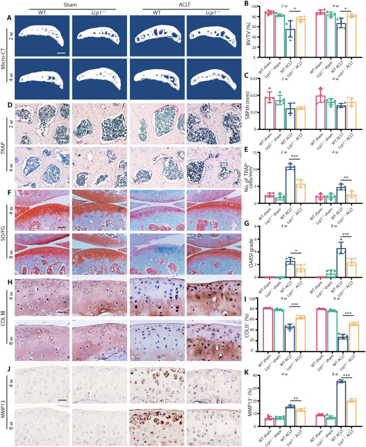
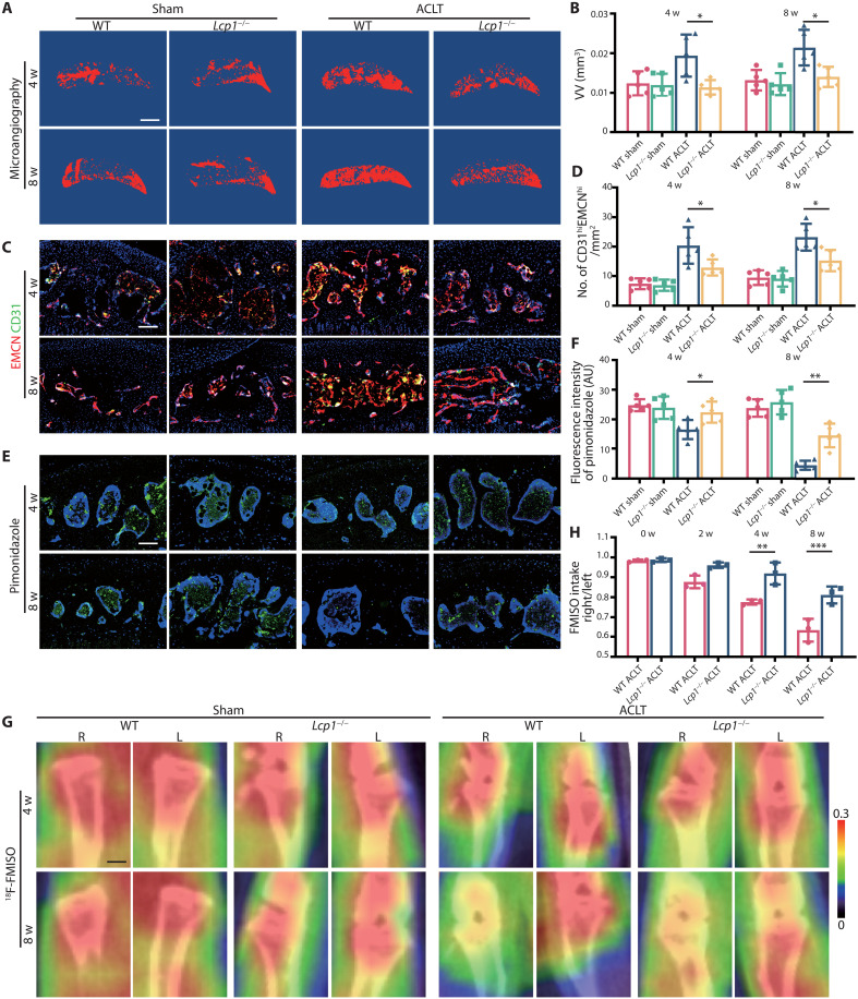
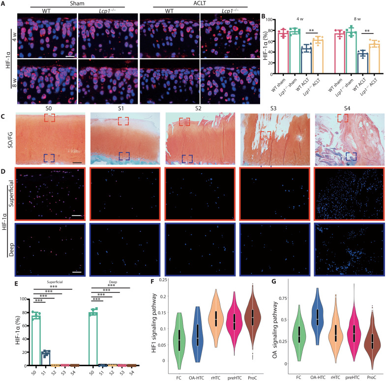
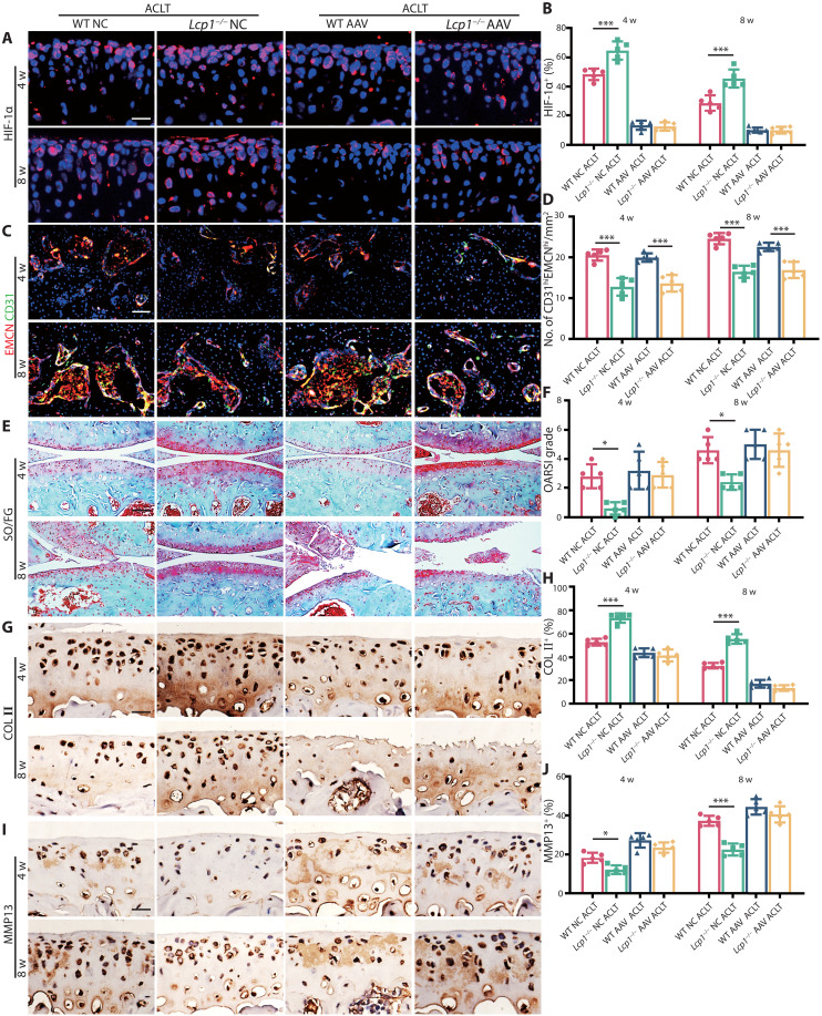
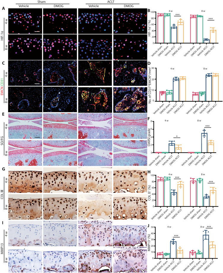
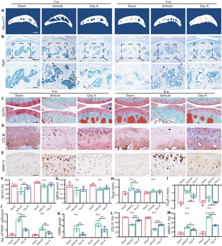
Abnormal subchondral bone remodeling featured by overactivated osteoclastogenesis leads to articular cartilage degeneration and osteoarthritis (OA) progression, but the mechanism is unclear. We used lymphocyte cytosolic protein 1 () knockout mice to suppress subchondral osteoclasts in a mice OA model with anterior cruciate ligament transection (ACLT), and mice showed decreased bone remodeling in subchondral bone and retarded cartilage degeneration. For mechanisms, the activated osteoclasts in subchondral bone induced type-H vessels and elevated oxygen concentration, which ubiquitylated hypoxia-inducible factor 1 alpha subunit (HIF-1α) in chondrocytes and led to cartilage degeneration. knockout impeded angiogenesis, which maintained hypoxia environment in joints and delayed the OA progression. Stabilization of HIF-1α delayed cartilage degeneration, and knockdown of abolished the protective effects of knockout. Last, we showed that Oroxylin A, an encoded protein l-plastin (LPL) inhibitor, could alleviate OA progression. In conclusion, maintaining hypoxic environment is an attractive strategy for OA treatment.
异常的软骨下骨重塑以破骨细胞过度激活为特征,导致关节软骨退化和骨关节炎(OA)进展,但机制尚不清楚。我们使用淋巴细胞胞浆蛋白 1()敲除小鼠抑制前交叉韧带切断(ACLT)的 OA 模型中的软骨下破骨细胞,结果 小鼠的软骨下骨骨重塑减少,软骨退化延迟。在机制上,软骨下骨中激活的破骨细胞诱导 H 型血管并提高氧浓度,从而导致软骨细胞中缺氧诱导因子 1α亚基(HIF-1α)泛素化,导致软骨退化。 敲除阻碍了血管生成,从而维持了关节中的低氧环境并延缓了 OA 的进展。HIF-1α 的稳定可延迟软骨退化,而 敲除则消除了 敲除的保护作用。最后,我们表明,白杨素 A,一种编码蛋白 l-肌动蛋白(LPL)抑制剂,可减轻 OA 的进展。总之,维持低氧环境是 OA 治疗的一种有吸引力的策略。