Zhai Aiguo, Peng Xuehua, Guo Yu, Li Jian, Shao Jianbo
Department of Imaging Center, Wuhan Children's Hospital (Wuhan Maternal and Child Healthcare Hospital), Tongji Medical College, Huazhong University of Science and Technology, Wuhan, China.
Front Pediatr. 2023 Mar 21;11:1089241. doi: 10.3389/fped.2023.1089241. eCollection 2023.
Our aim was to explore the clinical value of multimodal imaging examinations in the diagnosis of congenital pyriform fossa fistula in children, so as to provide clues for the early diagnosis and treatment of congenital pyriform fossa fistula.
The clinical and imaging data of 55 children with pyriform fossa fistula diagnosed surgically in our hospital from 2015 to 2018 were analyzed retrospectively. All 55 patients underwent a CT scan. Of those patients, contrast enhancement CT was performed in 47 cases, MRI was performed in 2 cases, and barium esophagography was performed in 41 cases.
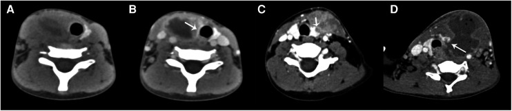
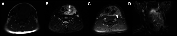
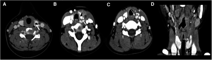
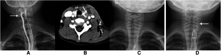
Among the 55 cases, there were 24 male patients and 31 female patients. The age ranged from 11 months to 13 years old, and the median age was 3.8 years old. The lesions of 49 cases (89.1%) were located on the left side, and the imaging of CT showed soft tissue mass in the anterior cervical region blurred boundary. There was ipsilateral thyroid involvement in 50 cases (90.9%), trachea and/or carotid sheath extension in 43 cases (78.2%), abscess formation in 39 cases (70.9%), and pneumatosis in 25 cases (45.5%). The CT examination of 22 children after treatment showed a linear or tubular low-density shadow in the thyroid gland, gas accumulation in the anterior cervical region or thyroid, and residual contrast medium, partly. A total of 24 cases underwent barium esophagography during the acute phase, and 15 cases (62.5%) showed sinus formation from the pyriform fossa downward or punctate high-density shadow in the anterior cervical region. The 2 cases where MRI was performed showed abscess formation in one side of the neck and thyroid involvement.
Pyriform fossa fistula is most common in the left anterior cervical region, and it is closely related to the thyroid gland. The plain and enhanced-contrast CT scan can be used as the first choice during the infection stage. It helps to understand the location, extent, and structure of the surrounding tissue. The preliminary diagnosis of pyriform sinus fistula was according to the imaging features. It provided an important basis for clinical diagnosis and reduced the pain caused by repeated infection or surgical incision and drainage.
探讨多模态影像学检查在儿童先天性梨状窝瘘诊断中的临床价值,为先天性梨状窝瘘的早期诊断与治疗提供线索。
回顾性分析2015年至2018年在我院手术确诊的55例梨状窝瘘患儿的临床及影像学资料。55例患者均行CT扫描,其中47例行增强CT扫描,2例行MRI检查,41例行食管钡餐造影。
55例中,男24例,女31例。年龄11个月至13岁,中位年龄3.8岁。49例(89.1%)病变位于左侧,CT影像表现为颈前区软组织肿块,边界不清。50例(90.9%)同侧甲状腺受累,43例(78.2%)气管和/或颈动脉鞘受累,39例(70.9%)形成脓肿,25例(45.5%)有积气。22例患儿治疗后CT检查显示甲状腺内有条状或管状低密度影,颈前区或甲状腺内有气体积聚,部分有残留造影剂。急性期共24例行食管钡餐造影,15例(62.5%)显示梨状窝向下形成窦道或颈前区点状高密度影。2例行MRI检查的病例显示颈部一侧形成脓肿且甲状腺受累。
梨状窝瘘最常见于颈前左侧,与甲状腺关系密切。平扫及增强CT扫描可作为感染期的首选检查,有助于了解病变部位、范围及周围组织的结构。根据影像学特征对梨状窦瘘进行初步诊断,为临床诊断提供重要依据,减少反复感染或手术切开引流带来的痛苦。