Physics Department, University of Pisa, Pisa, Italy.
Pisa Division, National Institute for Nuclear Physics, Pisa, Italy.
Eur Radiol Exp. 2023 Apr 10;7(1):18. doi: 10.1186/s41747-023-00334-z.
The role of computed tomography (CT) in the diagnosis and characterization of coronavirus disease 2019 (COVID-19) pneumonia has been widely recognized. We evaluated the performance of a software for quantitative analysis of chest CT, the LungQuant system, by comparing its results with independent visual evaluations by a group of 14 clinical experts. The aim of this work is to evaluate the ability of the automated tool to extract quantitative information from lung CT, relevant for the design of a diagnosis support model.
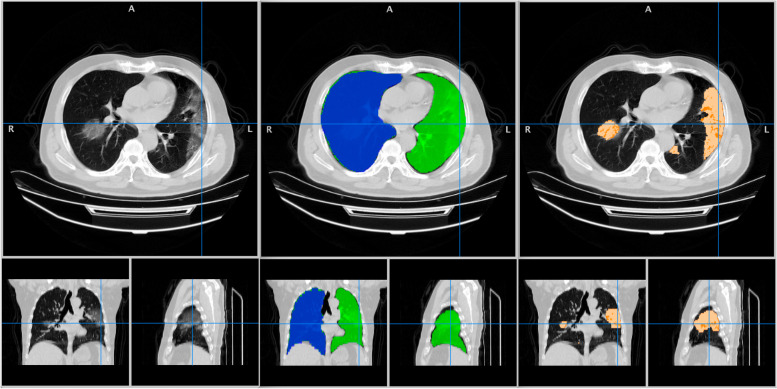
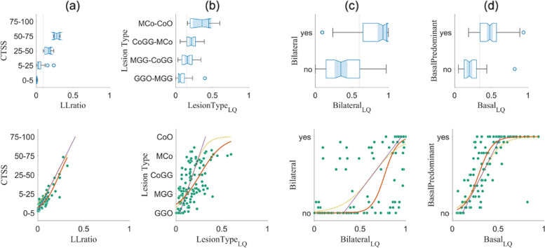
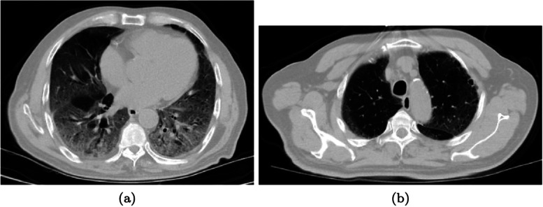
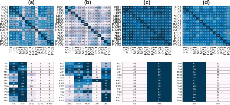
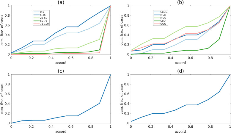
LungQuant segments both the lungs and lesions associated with COVID-19 pneumonia (ground-glass opacities and consolidations) and computes derived quantities corresponding to qualitative characteristics used to clinically assess COVID-19 lesions. The comparison was carried out on 120 publicly available CT scans of patients affected by COVID-19 pneumonia. Scans were scored for four qualitative metrics: percentage of lung involvement, type of lesion, and two disease distribution scores. We evaluated the agreement between the LungQuant output and the visual assessments through receiver operating characteristics area under the curve (AUC) analysis and by fitting a nonlinear regression model.
Despite the rather large heterogeneity in the qualitative labels assigned by the clinical experts for each metric, we found good agreement on the metrics compared to the LungQuant output. The AUC values obtained for the four qualitative metrics were 0.98, 0.85, 0.90, and 0.81.
Visual clinical evaluation could be complemented and supported by computer-aided quantification, whose values match the average evaluation of several independent clinical experts.
We conducted a multicenter evaluation of the deep learning-based LungQuant automated software. We translated qualitative assessments into quantifiable metrics to characterize coronavirus disease 2019 (COVID-19) pneumonia lesions. Comparing the software output to the clinical evaluations, results were satisfactory despite heterogeneity of the clinical evaluations. An automatic quantification tool may contribute to improve the clinical workflow of COVID-19 pneumonia.
计算机断层扫描(CT)在 2019 年冠状病毒病(COVID-19)肺炎的诊断和特征中的作用已得到广泛认可。我们通过将其结果与一组 14 位临床专家的独立视觉评估进行比较,评估了一种用于定量分析胸部 CT 的软件(LungQuant 系统)的性能。这项工作的目的是评估该自动工具从肺部 CT 中提取定量信息的能力,这对于设计诊断支持模型非常重要。
LungQuant 对 COVID-19 肺炎相关的肺部和病变(磨玻璃影和实变)进行分割,并计算与用于临床评估 COVID-19 病变的定性特征相对应的衍生量。比较是在 120 例公开的 COVID-19 肺炎患者 CT 扫描上进行的。对 4 种定性指标进行评分:肺受累百分比、病变类型和两种疾病分布评分。我们通过接受者操作特征曲线(AUC)分析和拟合非线性回归模型来评估 LungQuant 输出与视觉评估之间的一致性。
尽管临床专家为每个指标分配的定性标签存在较大差异,但与 LungQuant 输出相比,我们发现指标之间的一致性很好。四个定性指标的 AUC 值分别为 0.98、0.85、0.90 和 0.81。
视觉临床评估可以通过计算机辅助定量来补充和支持,其值与几个独立临床专家的平均评估相匹配。
我们对基于深度学习的 LungQuant 自动软件进行了多中心评估。我们将定性评估转化为可量化的指标,以对 COVID-19 肺炎病变进行特征描述。将软件输出与临床评估进行比较,结果令人满意,尽管临床评估存在异质性。自动定量工具可能有助于改善 COVID-19 肺炎的临床工作流程。