• 文献检索
  • 文档翻译
  • 深度研究
  • 学术资讯
  • Suppr Zotero 插件Zotero 插件
  • 邀请有礼
  • 套餐&价格
  • 历史记录
应用&插件
Suppr Zotero 插件Zotero 插件浏览器插件Mac 客户端Windows 客户端微信小程序
定价
高级版会员购买积分包购买API积分包
服务
文献检索文档翻译深度研究API 文档MCP 服务
关于我们
关于 Suppr公司介绍联系我们用户协议隐私条款
关注我们

Suppr 超能文献

核心技术专利:CN118964589B侵权必究
粤ICP备2023148730 号-1Suppr @ 2026

文献检索

告别复杂PubMed语法,用中文像聊天一样搜索,搜遍4000万医学文献。AI智能推荐,让科研检索更轻松。

立即免费搜索

文件翻译

保留排版,准确专业,支持PDF/Word/PPT等文件格式,支持 12+语言互译。

免费翻译文档

深度研究

AI帮你快速写综述,25分钟生成高质量综述,智能提取关键信息,辅助科研写作。

立即免费体验

肝癌中巨噬细胞 M0 相关基因特征的预后预测和多维剖析。

Prognostic prediction and multidimensional dissections of a macrophages M0-related gene signature in liver cancer.

机构信息

Department of Gastroenterology, Jining First People's Hospital, Jining, China.

Department of Radiotherapy Oncology, The Affiliated Yancheng First Hospital of Nanjing University Medical School, The First People's Hospital of Yancheng, Yancheng, China.

出版信息

Front Endocrinol (Lausanne). 2023 Mar 24;14:1153562. doi: 10.3389/fendo.2023.1153562. eCollection 2023.

DOI:10.3389/fendo.2023.1153562
PMID:37033261
原文链接:https://pmc.ncbi.nlm.nih.gov/articles/PMC10080084/
Abstract

BACKGROUND

Liver hepatocellular carcinoma (LIHC) is the seventh most commonly diagnosed malignancy and the third leading cause of all cancer death worldwide. The undifferentiated macrophages M0 can be induced into polarized M1 and M2 to exert opposite effects in tumor microenvironment. However, the prognostic value of macrophages M0 phenotype remains obscure in LIHC.

METHODS

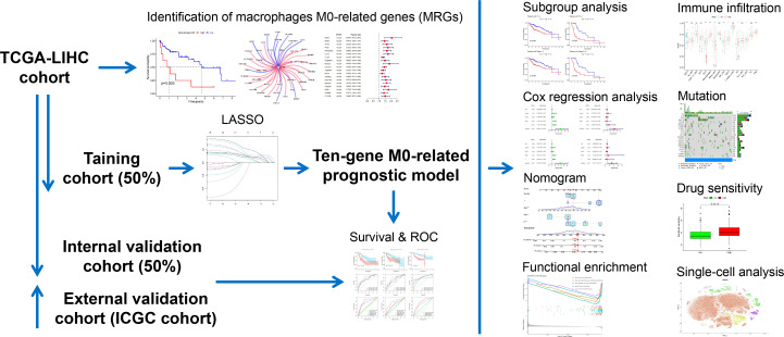
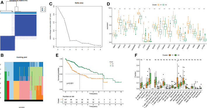
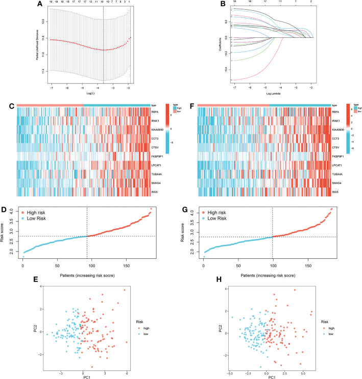
The transcriptome data of LIHC was obtained from TCGA database and ICGC database. 365 LIHC samples from TCGA database and 231 LIHC samples from ICGC database were finally included. Macrophages M0-related genes (MRGs) were screened by Pearson correlation analysis and univariate Cox regression analysis based on the infiltration level of Macrophages M0. LASSO regression analysis was employed to construct a prognostic signature based on MRGs, and risk scores were accordingly calculated. Then we investigated the MRGs-based prognostic signature with respects to prognostic value, clinical significance, strengthened pathways, immune infiltration, gene mutation and drug sensitivity. Furthermore, the expression pattern of MRGs in the tumor microenvironment were also detected in LIHC.

RESULTS

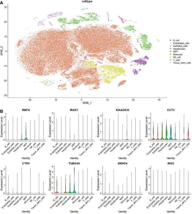
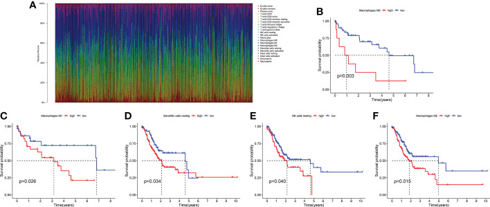
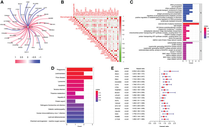
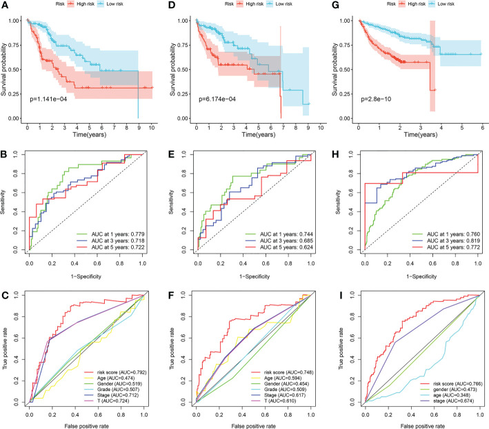
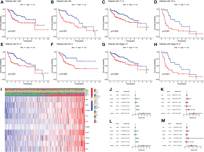
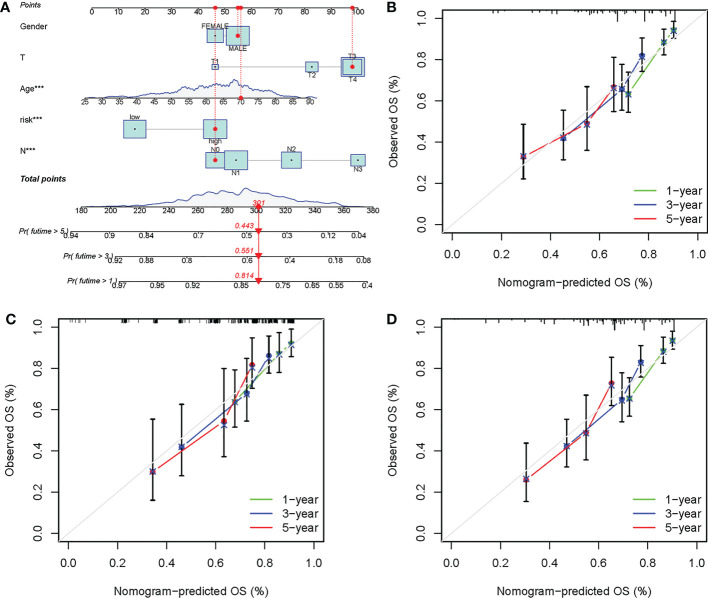
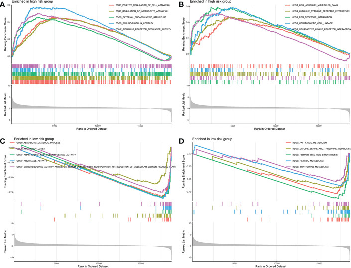
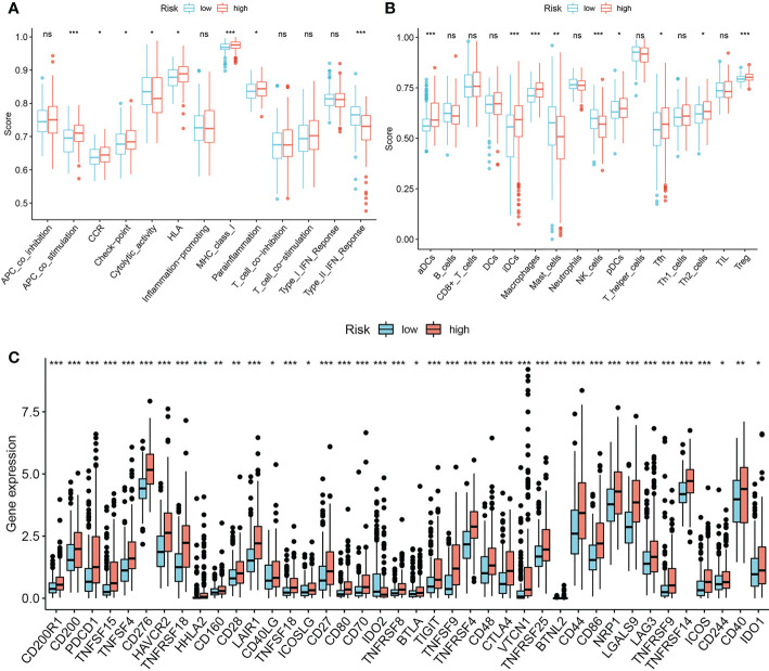
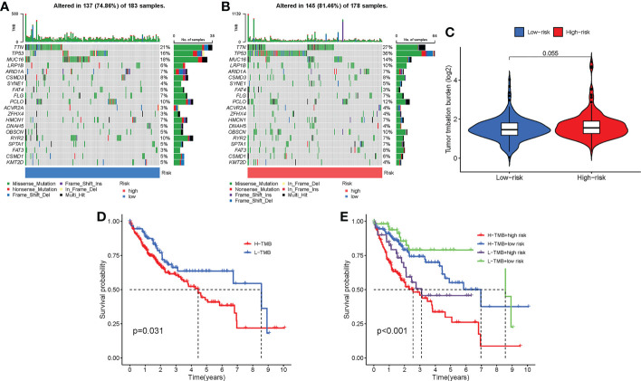
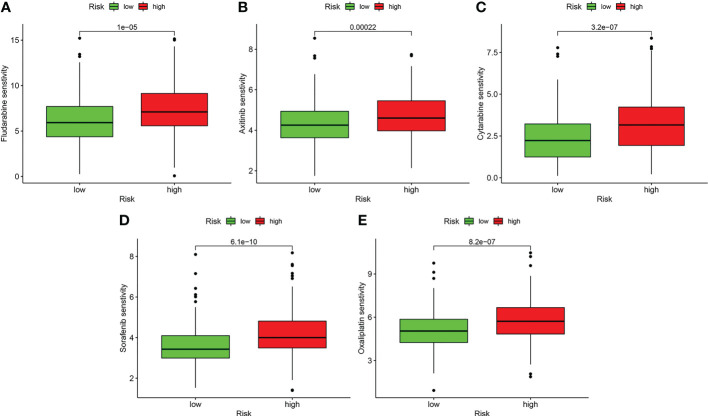
A ten-MRG signature was developed and clarified as independent prognostic predictors in LIHC. The risk score-based nomogram showed favorable capability in survival prediction. Several substance metabolism activities like fatty acid/amino acid metabolism were strengthened in low-risk group. Low risk group was deciphered to harbor TTN mutation-driven tumorigenesis, while TP53 mutation was dominant in high-risk group. We also ascertained that the infiltration levels of immune cells and expressions of immune checkpoints are significantly influenced by the risk score. Besides, we implied that patients in low-risk group may be more sensitive to several anti-cancer drugs. What's more important, single-cell analysis verified the expression of MRGs in the tumor microenvironment of LIHC.

CONCLUSION

Multidimensional evaluations verified the clinical utility of the macrophages M0-related gene signature to predict prognosis, assist risk decision and guide treatment strategy for patients with LIHC.

摘要

背景

肝肝细胞癌(LIHC)是全球第七种最常见的恶性肿瘤,也是所有癌症死亡的第三大主要原因。未分化的巨噬细胞 M0 可以被诱导为极化的 M1 和 M2,在肿瘤微环境中发挥相反的作用。然而,巨噬细胞 M0 表型在 LIHC 中的预后价值仍不清楚。

方法

从 TCGA 数据库和 ICGC 数据库中获取 LIHC 的转录组数据。最终纳入 TCGA 数据库的 365 个 LIHC 样本和 ICGC 数据库的 231 个 LIHC 样本。基于巨噬细胞 M0 的浸润水平,通过 Pearson 相关分析和单因素 Cox 回归分析筛选巨噬细胞 M0 相关基因(MRGs)。利用 LASSO 回归分析构建基于 MRGs 的预后特征,并据此计算风险评分。然后,我们从预后价值、临床意义、增强途径、免疫浸润、基因突变和药物敏感性等方面对基于 MRGs 的预后特征进行了研究。此外,还检测了 LIHC 肿瘤微环境中 MRGs 的表达模式。

结果

构建并验证了一个由十个 MRGs 组成的独立预后预测因子,用于 LIHC。基于风险评分的列线图在生存预测方面表现出良好的能力。在低风险组中,几种物质代谢活动(如脂肪酸/氨基酸代谢)得到了加强。低风险组揭示了 TTN 突变驱动的肿瘤发生,而高风险组则以 TP53 突变为主要特征。我们还确定了免疫细胞的浸润水平和免疫检查点的表达受到风险评分的显著影响。此外,我们暗示低风险组的患者可能对几种抗癌药物更敏感。更重要的是,单细胞分析验证了 LIHC 肿瘤微环境中 MRGs 的表达。

结论

多维度评估验证了巨噬细胞 M0 相关基因特征预测预后、辅助风险决策和指导 LIHC 患者治疗策略的临床实用性。

https://cdn.ncbi.nlm.nih.gov/pmc/blobs/406b/10080084/195634479dbf/fendo-14-1153562-g013.jpg
https://cdn.ncbi.nlm.nih.gov/pmc/blobs/406b/10080084/2e3e5ae048d3/fendo-14-1153562-g001.jpg
https://cdn.ncbi.nlm.nih.gov/pmc/blobs/406b/10080084/be32579ef3e0/fendo-14-1153562-g002.jpg
https://cdn.ncbi.nlm.nih.gov/pmc/blobs/406b/10080084/acd2d960cf73/fendo-14-1153562-g003.jpg
https://cdn.ncbi.nlm.nih.gov/pmc/blobs/406b/10080084/233c997fc74d/fendo-14-1153562-g004.jpg
https://cdn.ncbi.nlm.nih.gov/pmc/blobs/406b/10080084/2b26810410c3/fendo-14-1153562-g005.jpg
https://cdn.ncbi.nlm.nih.gov/pmc/blobs/406b/10080084/0290927af94e/fendo-14-1153562-g006.jpg
https://cdn.ncbi.nlm.nih.gov/pmc/blobs/406b/10080084/244342115ba8/fendo-14-1153562-g007.jpg
https://cdn.ncbi.nlm.nih.gov/pmc/blobs/406b/10080084/06f5378018b5/fendo-14-1153562-g008.jpg
https://cdn.ncbi.nlm.nih.gov/pmc/blobs/406b/10080084/954accc34c70/fendo-14-1153562-g009.jpg
https://cdn.ncbi.nlm.nih.gov/pmc/blobs/406b/10080084/9d88170278da/fendo-14-1153562-g010.jpg
https://cdn.ncbi.nlm.nih.gov/pmc/blobs/406b/10080084/9497102fb96c/fendo-14-1153562-g011.jpg
https://cdn.ncbi.nlm.nih.gov/pmc/blobs/406b/10080084/3896c119b9d0/fendo-14-1153562-g012.jpg
https://cdn.ncbi.nlm.nih.gov/pmc/blobs/406b/10080084/195634479dbf/fendo-14-1153562-g013.jpg

相似文献

1
Prognostic prediction and multidimensional dissections of a macrophages M0-related gene signature in liver cancer.肝癌中巨噬细胞 M0 相关基因特征的预后预测和多维剖析。
Front Endocrinol (Lausanne). 2023 Mar 24;14:1153562. doi: 10.3389/fendo.2023.1153562. eCollection 2023.
2
A novel signature incorporating lipid metabolism- and immune-related genes to predict the prognosis and immune landscape in hepatocellular carcinoma.一种包含脂质代谢和免疫相关基因的新型特征,用于预测肝细胞癌的预后和免疫格局。
Front Oncol. 2023 Jun 6;13:1182434. doi: 10.3389/fonc.2023.1182434. eCollection 2023.
3
Development of a macrophages-related 4-gene signature and nomogram for the overall survival prediction of hepatocellular carcinoma based on WGCNA and LASSO algorithm.基于 WGCNA 和 LASSO 算法构建巨噬细胞相关的 4 基因signature 及列线图预测肝细胞癌患者总生存期
Int Immunopharmacol. 2021 Jan;90:107238. doi: 10.1016/j.intimp.2020.107238. Epub 2020 Dec 11.
4
Development and validation of multi-omic prognostic signature of anoikis-related genes in liver hepatocellular carcinoma.多组学生存预后相关基因在肝肝细胞癌中的鉴定和验证。
Medicine (Baltimore). 2023 Nov 17;102(46):e36190. doi: 10.1097/MD.0000000000036190.
5
A signature based on neutrophil extracellular trap-related genes for the assessment of prognosis, immunoinfiltration, mutation and therapeutic response in hepatocellular carcinoma.基于中性粒细胞胞外诱捕网相关基因的signature 用于评估肝细胞癌的预后、免疫浸润、突变和治疗反应。
J Gene Med. 2024 Jan;26(1):e3588. doi: 10.1002/jgm.3588. Epub 2023 Sep 16.
6
A disulfidptosis-related lncRNAs signature in hepatocellular carcinoma: prognostic prediction, tumor immune microenvironment and drug susceptibility.肝细胞癌中与二硫键相关的 lncRNAs 特征:预后预测、肿瘤免疫微环境和药物敏感性。
Sci Rep. 2024 Jan 7;14(1):746. doi: 10.1038/s41598-024-51459-z.
7
A necroptosis-related gene signature for predicting prognosis, immune landscape, and drug sensitivity in hepatocellular carcinoma.一个与坏死性凋亡相关的基因特征,可用于预测肝细胞癌的预后、免疫图谱和药物敏感性。
Cancer Med. 2022 Dec;11(24):5079-5096. doi: 10.1002/cam4.4812. Epub 2022 May 13.
8
The fatty acid-related gene signature stratifies poor prognosis patients and characterizes TIME in cutaneous melanoma.脂肪酸相关基因特征对皮肤黑色素瘤预后不良患者进行分层,并对肿瘤免疫微环境进行表征。
J Cancer Res Clin Oncol. 2024 Jan 27;150(2):40. doi: 10.1007/s00432-023-05580-7.
9
Constructing and validating of m7G-related genes prognostic signature for hepatocellular carcinoma and immune infiltration: potential biomarkers for predicting the overall survival.构建和验证用于肝细胞癌的m7G相关基因预后特征及免疫浸润:预测总生存的潜在生物标志物
J Gastrointest Oncol. 2022 Dec;13(6):3169-3182. doi: 10.21037/jgo-22-1134.
10
Constructing immune and prognostic features associated with ADCP in hepatocellular carcinoma and pan-cancer based on scRNA-seq and bulk RNA-seq.基于 scRNA-seq 和 bulk RNA-seq 构建与 HCC 中 ADCP 相关的免疫和预后特征,并进行泛癌分析。
Front Immunol. 2024 May 7;15:1397541. doi: 10.3389/fimmu.2024.1397541. eCollection 2024.

引用本文的文献

1
The neutrophil extracellular trap-related gene FPR1 (formyl peptide receptor 1) as a potential prognostic and therapeutic target in osteosarcoma.中性粒细胞胞外诱捕网相关基因FPR1(甲酰肽受体1)作为骨肉瘤潜在的预后和治疗靶点。
BMC Musculoskelet Disord. 2025 Mar 31;26(1):309. doi: 10.1186/s12891-024-08231-1.
2
Integrative analysis of multi-omics data identified PLG as key gene related to Anoikis resistance and immune phenotypes in hepatocellular carcinoma.多组学数据的综合分析确定PLG是与肝细胞癌中失巢凋亡抗性和免疫表型相关的关键基因。
J Transl Med. 2024 Dec 4;22(1):1104. doi: 10.1186/s12967-024-05858-5.
3
Roles of M1 Macrophages and Their Extracellular Vesicles in Cancer Therapy.

本文引用的文献

1
An M0 macrophage-related prognostic model for hepatocellular carcinoma.M0 巨噬细胞相关的肝细胞癌预后模型。
BMC Cancer. 2022 Jul 19;22(1):791. doi: 10.1186/s12885-022-09872-y.
2
T-cell exhaustion and residency dynamics inform clinical outcomes in hepatocellular carcinoma.T 细胞耗竭和驻留动力学为肝细胞癌的临床结局提供信息。
J Hepatol. 2022 Aug;77(2):397-409. doi: 10.1016/j.jhep.2022.02.032. Epub 2022 Apr 1.
3
Tumor-associated macrophages related signature in glioma.肿瘤相关巨噬细胞相关特征在神经胶质瘤中的表现。
M1 巨噬细胞及其细胞外囊泡在癌症治疗中的作用。
Cells. 2024 Aug 26;13(17):1428. doi: 10.3390/cells13171428.
4
Matrix Metalloproteinase 11 Promotes Migration and Invasion of Colorectal Cancer by Elevating Slug Protein.基质金属蛋白酶 11 通过升高 Slug 蛋白促进结直肠癌的迁移和侵袭。
Int J Med Sci. 2024 Aug 13;21(11):2170-2188. doi: 10.7150/ijms.98007. eCollection 2024.
5
Identification and validation of immune-related gene signature models for predicting prognosis and immunotherapy response in hepatocellular carcinoma.鉴定和验证免疫相关基因特征模型,以预测肝细胞癌的预后和免疫治疗反应。
Front Immunol. 2024 Jun 12;15:1371829. doi: 10.3389/fimmu.2024.1371829. eCollection 2024.
Aging (Albany NY). 2022 Mar 24;14(6):2720-2735. doi: 10.18632/aging.203968.
4
Metabolic dysregulation and emerging therapeutical targets for hepatocellular carcinoma.肝细胞癌的代谢失调与新兴治疗靶点
Acta Pharm Sin B. 2022 Feb;12(2):558-580. doi: 10.1016/j.apsb.2021.09.019. Epub 2021 Sep 25.
5
Immunotherapies for hepatocellular carcinoma.肝细胞癌的免疫疗法
Nat Rev Clin Oncol. 2022 Mar;19(3):151-172. doi: 10.1038/s41571-021-00573-2. Epub 2021 Nov 11.
6
Clinical M2 macrophages-related genes to aid therapy in pancreatic ductal adenocarcinoma.用于辅助治疗胰腺导管腺癌的临床M2巨噬细胞相关基因。
Cancer Cell Int. 2021 Oct 30;21(1):582. doi: 10.1186/s12935-021-02289-w.
7
Hepatocellular Carcinoma Immunotherapy.肝细胞癌免疫疗法
Annu Rev Med. 2022 Jan 27;73:267-278. doi: 10.1146/annurev-med-042220-021121. Epub 2021 Oct 4.
8
Advances in immunotherapy for hepatocellular carcinoma.肝细胞癌的免疫治疗进展。
Nat Rev Gastroenterol Hepatol. 2021 Aug;18(8):525-543. doi: 10.1038/s41575-021-00438-0. Epub 2021 Apr 13.
9
Global Cancer Statistics 2020: GLOBOCAN Estimates of Incidence and Mortality Worldwide for 36 Cancers in 185 Countries.《全球癌症统计数据 2020:全球 185 个国家和地区 36 种癌症的发病率和死亡率估计》。
CA Cancer J Clin. 2021 May;71(3):209-249. doi: 10.3322/caac.21660. Epub 2021 Feb 4.
10
Single-cell landscape of the ecosystem in early-relapse hepatocellular carcinoma.早期复发肝细胞癌生态系统的单细胞景观。
Cell. 2021 Jan 21;184(2):404-421.e16. doi: 10.1016/j.cell.2020.11.041. Epub 2020 Dec 23.