Department of Biomedical, Surgical and Dental Sciences, University of Milan, Milan, Italy.
Foundation IRCCS Ca' Granda Polyclinic, Via Della Commenda 10, 20122, Milan, Italy.
Clin Oral Investig. 2023 Jul;27(7):3779-3786. doi: 10.1007/s00784-023-04995-3. Epub 2023 Apr 13.
The relationship between the anatomy of the interradicular space and success in regenerative therapy of furcation defects is discussed in this paper. The goal of this retrospective, multicenter clinical study is to clinically evaluate the relationship between the interradicular conformation and regenerative therapy success with the use of a novel measurement method.
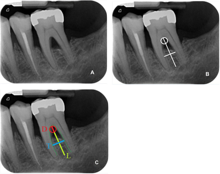
One hundred thirty-eight radiographs of mandibular molars with furcation defects that had been treated with regenerative therapy were collected from six clinical centers. Data on the type of therapy and clinical parameters before and after treatment (follow-up of at least 12 months) were collected. The radiographs (before surgery and at least 12 months postoperatively) were measured with a visual evaluation method by a blind operator using graphics software.
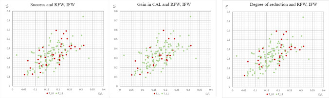
Success, defined as a reduction in horizontal and vertical furcation involvement, decrease in probing depths, and increase in clinical attachment level, was statistically assessed on 138 regenerated molars sites and were related to clinical variables such as age, sex, center, and treatment. No correlation was found between success in regenerative therapy and the conformation of the interradicular space, measured with a visual ratio method and a standard linear measurement. At the univariate analysis, the parameters that had a correlation with success were center, extent of furcation involvement, treatment, and sex. The use of enamel matrix derivative (EMD) seemed to be the most favorable therapy, with increase in CAL gain and reduction of vertical or horizontal furcation involvement.
The regenerative outcome was not significantly influenced by the anatomy of furcation. The center, the degree of furcation involvement, sex, and treatment (EMD) were significantly associated with higher success of periodontal regeneration.
本文讨论了根间空间解剖结构与分叉缺损再生治疗成功之间的关系。本回顾性、多中心临床研究的目的是临床评估根间形态与使用新型测量方法的再生治疗成功之间的关系。
从六个临床中心收集了 138 张下颌磨牙分叉缺损经再生治疗的射线照片。收集了治疗类型和治疗前后(至少 12 个月的随访)临床参数的数据。使用图形软件,由一名盲操作者通过视觉评估方法对射线照片(术前和至少 12 个月的术后)进行测量。
成功定义为水平和垂直分叉受累减少、探诊深度减小和临床附着水平增加,对 138 个再生磨牙部位进行了统计学评估,并与年龄、性别、中心和治疗等临床变量相关。用视觉比法和标准线性测量法测量根间空间形态与再生治疗的成功之间未发现相关性。在单变量分析中,与成功相关的参数为中心、分叉受累程度、治疗和性别。使用釉基质衍生物(EMD)似乎是最有利的治疗方法,可增加 CAL 增益并减少垂直或水平分叉受累。
再生治疗结果不受分叉解剖结构的显著影响。中心、分叉受累程度、性别和治疗(EMD)与牙周再生更高的成功率显著相关。