• 文献检索
  • 文档翻译
  • 深度研究
  • 学术资讯
  • Suppr Zotero 插件Zotero 插件
  • 邀请有礼
  • 套餐&价格
  • 历史记录
应用&插件
Suppr Zotero 插件Zotero 插件浏览器插件Mac 客户端Windows 客户端微信小程序
定价
高级版会员购买积分包购买API积分包
服务
文献检索文档翻译深度研究API 文档MCP 服务
关于我们
关于 Suppr公司介绍联系我们用户协议隐私条款
关注我们

Suppr 超能文献

核心技术专利:CN118964589B侵权必究
粤ICP备2023148730 号-1Suppr @ 2026

文献检索

告别复杂PubMed语法,用中文像聊天一样搜索,搜遍4000万医学文献。AI智能推荐,让科研检索更轻松。

立即免费搜索

文件翻译

保留排版,准确专业,支持PDF/Word/PPT等文件格式,支持 12+语言互译。

免费翻译文档

深度研究

AI帮你快速写综述,25分钟生成高质量综述,智能提取关键信息,辅助科研写作。

立即免费体验

创伤性脑损伤模型的特征:从宏观血流变化到微观线粒体变化

Characteristics of traumatic brain injury models: from macroscopic blood flow changes to microscopic mitochondrial changes.

作者信息

Yang Ding-Ding, Wan Xiang-Dong, Chen An-Di, Yan Zi-Qian, Lu Yi-Fan, Liu Jun-Chen, Wang Ya-Zhou, Wang Jing, Zhao Yan, Wu Sheng-Xi, Cai Guo-Hong

机构信息

Department of Neurobiology, School of Basic Medicine, Forth Military Medical University, Xi'an, Shaanxi Province, China.

Department of Neurobiology, School of Basic Medicine, Forth Military Medical University, Xi'an, Shaanxi Province; Hebei Medical University, Shijiazhuang, Hebei Province, China.

出版信息

Neural Regen Res. 2023 Oct;18(10):2268-2277. doi: 10.4103/1673-5374.369125.

DOI:10.4103/1673-5374.369125
PMID:37056147
原文链接:https://pmc.ncbi.nlm.nih.gov/articles/PMC10328283/
Abstract

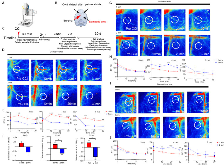
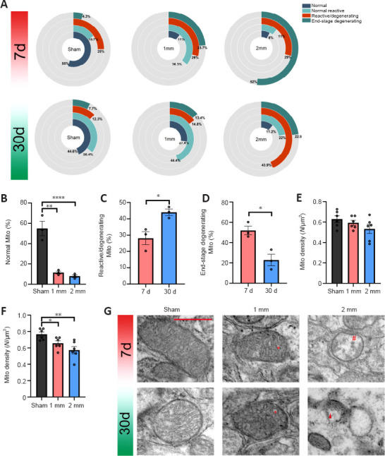
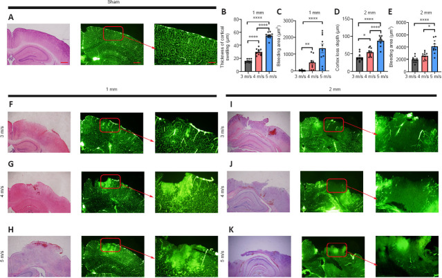
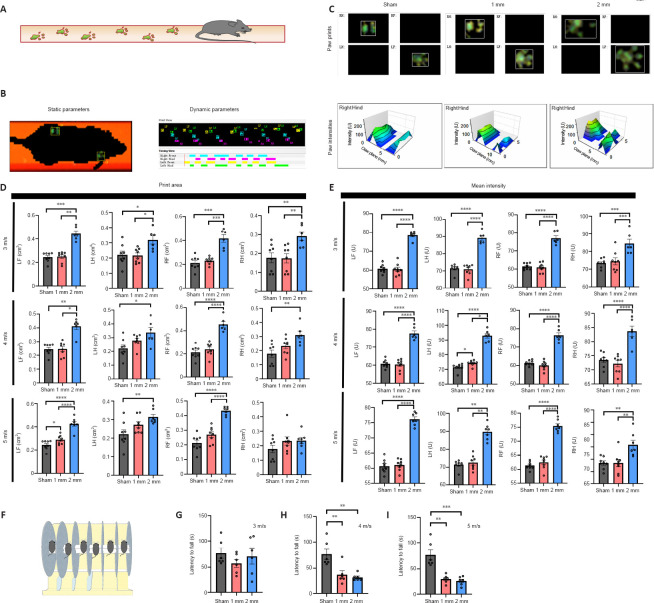
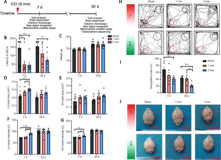
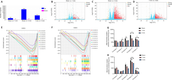
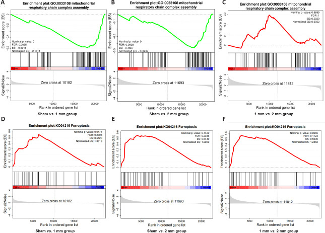
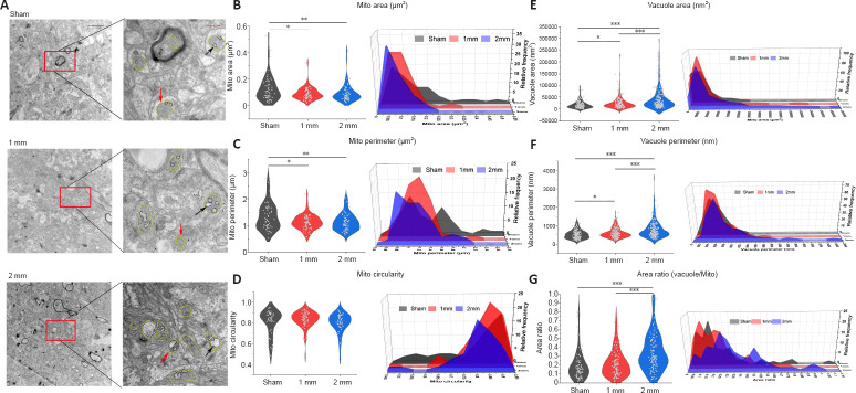
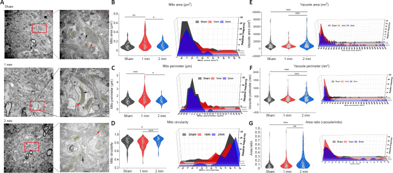
Controlled cortical impingement is a widely accepted method to induce traumatic brain injury to establish a traumatic brain injury animal model. A strike depth of 1 mm at a certain speed is recommended for a moderate brain injury and a depth of > 2 mm is used to induce severe brain injury. However, the different effects and underlying mechanisms of these two model types have not been proven. This study investigated the changes in cerebral blood flow, differences in the degree of cortical damage, and differences in motor function under different injury parameters of 1 and 2 mm at injury speeds of 3, 4, and 5 m/s. We also explored the functional changes and mitochondrial damage between the 1 and 2 mm groups in the acute (7 days) and chronic phases (30 days). The results showed that the cerebral blood flow in the injured area of the 1 mm group was significantly increased, and swelling and bulging of brain tissue, increased vascular permeability, and large-scale exudation occurred. In the 2 mm group, the main pathological changes were decreased cerebral blood flow, brain tissue loss, and cerebral vasospasm occlusion in the injured area. Substantial motor and cognitive impairments were found on day 7 after injury in the 2 mm group; at 30 days after injury, the motor function of the 2 mm group mice recovered significantly while cognitive impairment persisted. Transcriptome sequencing showed that compared with the 1 mm group, the 2 mm group expressed more ferroptosis-related genes. Morphological changes of mitochondria in the two groups on days 7 and 30 using transmission electron microscopy revealed that on day 7, the mitochondria in both groups shrank and the vacuoles became larger; on day 30, the mitochondria in the 1 mm group became larger, and the vacuoles in the 2 mm group remained enlarged. By analyzing the proportion of mitochondrial subgroups in different groups, we found that the model mice had different patterns of mitochondrial composition at different time periods, suggesting that the difference in the degree of damage among traumatic brain injury groups may reflect the mitochondrial changes. Taken together, differences in mitochondrial morphology and function between the 1 and 2 mm groups provide a new direction for the accurate classification of traumatic brain injury. Our results provide reliable data support and evaluation methods for promoting the establishment of standard mouse controlled cortical impingement model guidelines.

摘要

控制性皮质撞击是一种广泛接受的诱导创伤性脑损伤以建立创伤性脑损伤动物模型的方法。对于中度脑损伤,建议以一定速度撞击深度为1毫米,而撞击深度>2毫米则用于诱导重度脑损伤。然而,这两种模型类型的不同影响及潜在机制尚未得到证实。本研究调查了在3、4和5米/秒的损伤速度下,1毫米和2毫米不同损伤参数时脑血流量的变化、皮质损伤程度的差异以及运动功能的差异。我们还探究了急性(7天)和慢性期(30天)1毫米和2毫米组之间的功能变化和线粒体损伤。结果显示,1毫米组损伤区域的脑血流量显著增加,脑组织出现肿胀和膨出,血管通透性增加以及大量渗出。在2毫米组中,主要病理变化为损伤区域脑血流量减少、脑组织缺失以及脑血管痉挛闭塞。在损伤后第7天,2毫米组发现明显的运动和认知障碍;在损伤后30天,2毫米组小鼠的运动功能显著恢复,而认知障碍持续存在。转录组测序显示,与1毫米组相比,2毫米组表达更多铁死亡相关基因。使用透射电子显微镜观察两组在第7天和第30天的线粒体形态变化,结果显示在第7天,两组线粒体均萎缩且空泡变大;在第30天,1毫米组线粒体变大,而2毫米组空泡仍扩大。通过分析不同组中线粒体亚群的比例,我们发现模型小鼠在不同时间段具有不同的线粒体组成模式,这表明创伤性脑损伤组之间损伤程度的差异可能反映了线粒体变化。综上所述,1毫米和2毫米组之间线粒体形态和功能的差异为创伤性脑损伤的准确分类提供了新方向。我们的结果为促进标准小鼠控制性皮质撞击模型指南的建立提供了可靠的数据支持和评估方法。

https://cdn.ncbi.nlm.nih.gov/pmc/blobs/37c3/10328283/a577d8fb36dd/NRR-18-2268-g010.jpg
https://cdn.ncbi.nlm.nih.gov/pmc/blobs/37c3/10328283/a7a75275ca13/NRR-18-2268-g002.jpg
https://cdn.ncbi.nlm.nih.gov/pmc/blobs/37c3/10328283/d6620e2569f0/NRR-18-2268-g003.jpg
https://cdn.ncbi.nlm.nih.gov/pmc/blobs/37c3/10328283/80daad739269/NRR-18-2268-g004.jpg
https://cdn.ncbi.nlm.nih.gov/pmc/blobs/37c3/10328283/fc8c8ae25950/NRR-18-2268-g005.jpg
https://cdn.ncbi.nlm.nih.gov/pmc/blobs/37c3/10328283/1e2a25317160/NRR-18-2268-g006.jpg
https://cdn.ncbi.nlm.nih.gov/pmc/blobs/37c3/10328283/0c9e5a3fa2f4/NRR-18-2268-g007.jpg
https://cdn.ncbi.nlm.nih.gov/pmc/blobs/37c3/10328283/0d323e9d8bb5/NRR-18-2268-g008.jpg
https://cdn.ncbi.nlm.nih.gov/pmc/blobs/37c3/10328283/fc62b0b2fc20/NRR-18-2268-g009.jpg
https://cdn.ncbi.nlm.nih.gov/pmc/blobs/37c3/10328283/a577d8fb36dd/NRR-18-2268-g010.jpg

相似文献

1
Characteristics of traumatic brain injury models: from macroscopic blood flow changes to microscopic mitochondrial changes.创伤性脑损伤模型的特征:从宏观血流变化到微观线粒体变化
Neural Regen Res. 2023 Oct;18(10):2268-2277. doi: 10.4103/1673-5374.369125.
2
Differences in pathological changes between two rat models of severe traumatic brain injury.两种重度创伤性脑损伤大鼠模型的病理变化差异。
Neural Regen Res. 2019 Oct;14(10):1796-1804. doi: 10.4103/1673-5374.257534.
3
Reversible Disruption of Neuronal Mitochondria by Ischemic and Traumatic Injury Revealed by Quantitative Two-Photon Imaging in the Neocortex of Anesthetized Mice.定量双光子成像揭示麻醉小鼠新皮质中缺血和创伤性损伤对神经元线粒体的可逆性破坏
J Neurosci. 2017 Jan 11;37(2):333-348. doi: 10.1523/JNEUROSCI.1510-16.2016.
4
Time course of post-traumatic mitochondrial oxidative damage and dysfunction in a mouse model of focal traumatic brain injury: implications for neuroprotective therapy.局灶性创伤性脑损伤小鼠模型中创伤后线粒体氧化损伤和功能障碍的时间进程:对神经保护治疗的意义。
J Cereb Blood Flow Metab. 2006 Nov;26(11):1407-18. doi: 10.1038/sj.jcbfm.9600297. Epub 2006 Mar 15.
5
Inhibition of ferroptosis attenuates tissue damage and improves long-term outcomes after traumatic brain injury in mice.铁死亡抑制减轻了小鼠创伤性脑损伤后的组织损伤并改善了长期预后。
CNS Neurosci Ther. 2019 Apr;25(4):465-475. doi: 10.1111/cns.13069. Epub 2018 Sep 28.
6
Acute, transient hemorrhagic hypotension does not aggravate structural damage or neurologic motor deficits but delays the long-term cognitive recovery following mild to moderate traumatic brain injury.急性短暂性出血性低血压不会加重结构损伤或神经运动功能缺损,但会延迟轻度至中度创伤性脑损伤后的长期认知恢复。
Crit Care Med. 2006 Feb;34(2):492-501. doi: 10.1097/01.ccm.0000198326.32049.7f.
7
Structural and functional damage sustained by mitochondria after traumatic brain injury in the rat: evidence for differentially sensitive populations in the cortex and hippocampus.大鼠创伤性脑损伤后线粒体的结构和功能损伤:皮层和海马中不同敏感群体的证据。
J Cereb Blood Flow Metab. 2003 Feb;23(2):219-31. doi: 10.1097/01.WCB.0000040581.43808.03.
8
Glutamate receptor antibodies in neurological diseases: anti-AMPA-GluR3 antibodies, anti-NMDA-NR1 antibodies, anti-NMDA-NR2A/B antibodies, anti-mGluR1 antibodies or anti-mGluR5 antibodies are present in subpopulations of patients with either: epilepsy, encephalitis, cerebellar ataxia, systemic lupus erythematosus (SLE) and neuropsychiatric SLE, Sjogren's syndrome, schizophrenia, mania or stroke. These autoimmune anti-glutamate receptor antibodies can bind neurons in few brain regions, activate glutamate receptors, decrease glutamate receptor's expression, impair glutamate-induced signaling and function, activate blood brain barrier endothelial cells, kill neurons, damage the brain, induce behavioral/psychiatric/cognitive abnormalities and ataxia in animal models, and can be removed or silenced in some patients by immunotherapy.神经疾病中的谷氨酸受体抗体:抗AMPA - GluR3抗体、抗NMDA - NR1抗体、抗NMDA - NR2A/B抗体、抗mGluR1抗体或抗mGluR5抗体存在于以下疾病患者的亚组中:癫痫、脑炎、小脑共济失调、系统性红斑狼疮(SLE)和神经精神性SLE、干燥综合征、精神分裂症、躁狂症或中风。这些自身免疫性抗谷氨酸受体抗体可在少数脑区与神经元结合,激活谷氨酸受体,降低谷氨酸受体的表达,损害谷氨酸诱导的信号传导和功能,激活血脑屏障内皮细胞,杀死神经元,损伤大脑,在动物模型中诱发行为/精神/认知异常和共济失调,并且在一些患者中可通过免疫疗法去除或使其失活。
J Neural Transm (Vienna). 2014 Aug;121(8):1029-75. doi: 10.1007/s00702-014-1193-3. Epub 2014 Aug 1.
9
Temporal changes in inflammatory mitochondria-enriched microRNAs following traumatic brain injury and effects of miR-146a nanoparticle delivery.创伤性脑损伤后富含炎症线粒体的微小RNA的时间变化及miR-146a纳米颗粒递送的影响
Neural Regen Res. 2021 Mar;16(3):514-522. doi: 10.4103/1673-5374.293149.
10
Mitochondrial bioenergetic alterations after focal traumatic brain injury in the immature brain.未成熟脑局灶性创伤性脑损伤后的线粒体生物能量改变
Exp Neurol. 2015 Sep;271:136-44. doi: 10.1016/j.expneurol.2015.05.009. Epub 2015 May 28.

引用本文的文献

1
Topological functional network analysis of cortical blood flow in hyperacute ischemic rats.超急性缺血大鼠皮质血流的拓扑功能网络分析
Brain Struct Funct. 2024 Dec 26;230(1):20. doi: 10.1007/s00429-024-02864-7.
2
Biomaterials and tissue engineering in traumatic brain injury: novel perspectives on promoting neural regeneration.创伤性脑损伤中的生物材料与组织工程:促进神经再生的新视角
Neural Regen Res. 2024 Oct 1;19(10):2157-2174. doi: 10.4103/1673-5374.391179. Epub 2023 Dec 21.

本文引用的文献

1
Spatial-temporal changes of iron deposition and iron metabolism after traumatic brain injury in mice.小鼠创伤性脑损伤后铁沉积及铁代谢的时空变化
Front Mol Neurosci. 2022 Aug 11;15:949573. doi: 10.3389/fnmol.2022.949573. eCollection 2022.
2
Mitochondria-Related Ferroptosis Drives Cognitive Deficits in Neonatal Mice Following Sevoflurane Administration.线粒体相关的铁死亡驱动新生小鼠在七氟醚给药后出现认知缺陷。
Front Med (Lausanne). 2022 Jul 22;9:887062. doi: 10.3389/fmed.2022.887062. eCollection 2022.
3
Risk and cause of death in post-traumatic epilepsy: a register-based retrospective cohort study.
创伤后癫痫的风险和死因:基于登记的回顾性队列研究。
J Neurol. 2022 Nov;269(11):6014-6020. doi: 10.1007/s00415-022-11279-5. Epub 2022 Jul 19.
4
Discovery of Notch Pathway-Related Genes for Predicting Prognosis and Tumor Microenvironment Status in Bladder Cancer.发现用于预测膀胱癌预后和肿瘤微环境状态的Notch信号通路相关基因
Front Genet. 2022 Jun 30;13:928778. doi: 10.3389/fgene.2022.928778. eCollection 2022.
5
Cerebral venous hemodynamic responses in a mouse model of traumatic brain injury.创伤性脑损伤小鼠模型中的脑静脉血液动力学反应。
Brain Res. 2022 Oct 1;1792:148014. doi: 10.1016/j.brainres.2022.148014. Epub 2022 Jul 12.
6
The Effect of Cerebrolysin on Anxiety, Depression, and Cognition in Moderate and Severe Traumatic Brain Injury Patients: A CAPTAIN II Retrospective Trial Analysis.脑活素对中重度创伤性脑损伤患者焦虑、抑郁和认知功能的影响:CAPTAIN II 回顾性试验分析。
Medicina (Kaunas). 2022 May 9;58(5):648. doi: 10.3390/medicina58050648.
7
Early Cerebral Blood Flow Changes, Cerebrovascular Reactivity and Cortical Spreading Depolarizations in Experimental Mild Traumatic Brain Injury Model.实验性轻度创伤性脑损伤模型中的早期脑血流变化、脑血管反应性和皮质扩散性去极化
Turk Neurosurg. 2022;32(3):488-499. doi: 10.5137/1019-5149.JTN.37742-22.1.
8
The prevalence and risk factors of posttraumatic cerebral infarction in patients with traumatic brain injury: a systematic review and meta-analysis.颅脑创伤患者创伤后脑梗死的患病率及相关危险因素:系统评价和荟萃分析。
Bioengineered. 2022 May;13(5):11706-11717. doi: 10.1080/21655979.2022.2070999.
9
Theoretical analysis of reversible and irreversible mitochondrial swelling invivo.体内可逆和不可逆线粒体肿胀的理论分析。
Biosystems. 2022 Jul;217:104679. doi: 10.1016/j.biosystems.2022.104679. Epub 2022 Apr 9.
10
Individuals with Chronic Mild-to-Moderate Traumatic Brain Injury Exhibit Decreased Neuromuscular Complexity During Gait.患有慢性轻度至中度创伤性脑损伤的个体在步态过程中表现出神经肌肉复杂性降低。
Neurorehabil Neural Repair. 2022 Apr;36(4-5):317-327. doi: 10.1177/15459683221081064. Epub 2022 Mar 23.