Li Xin, Su Fengbo, Yuan Qinghai, Chen Yan, Liu Chun-Yan, Fan Yuhang
Department of Radiology, the Second Hospital of Jilin University, Changchun, China.
Department of Neurosurgery, the Second Hospital of Jilin University, Changchun, China.
Quant Imaging Med Surg. 2023 Apr 1;13(4):2712-2734. doi: 10.21037/qims-22-750. Epub 2023 Feb 22.
Cerebrovascular diseases (CVDs), particularly cerebral stroke, remain a primary cause of disability and death worldwide. Accurate diagnosis of CVDs is essential to guide therapeutic decisions and foresee the prognosis. Different CVDs have different pathological processes while they have many signs in common with some other brain diseases. Thus, differential diagnoses of strokes from other primary and secondary CVDs are especially important and challenging.
This review is composed mainly based on searching PubMed articles between September, 2013 and December 26, 2022 in English.
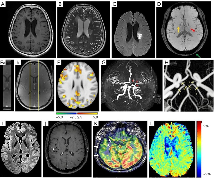
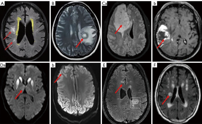
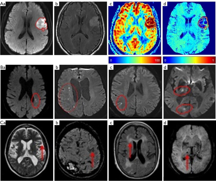
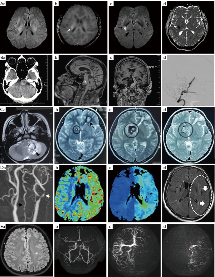
Neuroimaging is a powerful tool for CVD diagnosis including cerebral angiography, ultrasound, computed tomography, and positron emission tomography as well as magnetic resonance imaging (MRI). MRI excels other imaging techniques by its features of non-invasive, diverse sequences and high spatiotemporal resolution. It can detect hemodynamic, structural alterations of intracranial arteries and metabolic status of their associated brain regions. In acute stroke, differential diagnosis of ischemic from hemorrhagic stroke and other intracranial vasculopathies is a common application of MRI. By providing information about the pathological characteristics of cerebral diseases exhibiting different degrees of behavioral alterations, cognitive impairment, motor dysfunction and other indications, MRI can differentiate strokes from other primary CVDs involving cerebral small vessels and identify vascular dementia from hyponatremia, brain tumors and other secondary or non-primary CVDs.
Recent advances in MRI technology allow clinical neuroimaging to provide unique reference for differentiating many previously inconclusive CVDs. MRI technology is worthy of full exploration while breaking its limitations in clinical applications should be considered.
脑血管疾病(CVDs),尤其是脑卒中,仍是全球致残和致死的主要原因。准确诊断CVDs对于指导治疗决策和预测预后至关重要。不同的CVDs有不同的病理过程,同时它们与其他一些脑部疾病有许多共同症状。因此,将脑卒中与其他原发性和继发性CVDs进行鉴别诊断尤为重要且具有挑战性。
本综述主要基于检索2013年9月至2022年12月26日期间的英文PubMed文章撰写而成。
神经影像学是诊断CVDs的有力工具,包括脑血管造影、超声、计算机断层扫描、正电子发射断层扫描以及磁共振成像(MRI)。MRI凭借其无创、序列多样和高时空分辨率的特点优于其他成像技术。它可以检测颅内动脉的血流动力学、结构改变及其相关脑区的代谢状态。在急性脑卒中中,鉴别缺血性脑卒中与出血性脑卒中及其他颅内血管病变是MRI的常见应用。通过提供有关表现出不同程度行为改变、认知障碍、运动功能障碍及其他症状的脑部疾病病理特征的信息,MRI可以将脑卒中与其他累及脑小血管的原发性CVDs区分开来,并将血管性痴呆与低钠血症、脑肿瘤及其他继发性或非原发性CVDs区分开来。
MRI技术的最新进展使临床神经影像学能够为鉴别许多以前无法确诊的CVDs提供独特的参考依据。MRI技术值得充分探索,同时应考虑突破其在临床应用中的局限性。