• 文献检索
  • 文档翻译
  • 深度研究
  • 学术资讯
  • Suppr Zotero 插件Zotero 插件
  • 邀请有礼
  • 套餐&价格
  • 历史记录
应用&插件
Suppr Zotero 插件Zotero 插件浏览器插件Mac 客户端Windows 客户端微信小程序
定价
高级版会员购买积分包购买API积分包
服务
文献检索文档翻译深度研究API 文档MCP 服务
关于我们
关于 Suppr公司介绍联系我们用户协议隐私条款
关注我们

Suppr 超能文献

核心技术专利:CN118964589B侵权必究
粤ICP备2023148730 号-1Suppr @ 2026

文献检索

告别复杂PubMed语法,用中文像聊天一样搜索,搜遍4000万医学文献。AI智能推荐,让科研检索更轻松。

立即免费搜索

文件翻译

保留排版,准确专业,支持PDF/Word/PPT等文件格式,支持 12+语言互译。

免费翻译文档

深度研究

AI帮你快速写综述,25分钟生成高质量综述,智能提取关键信息,辅助科研写作。

立即免费体验

靶向内皮细胞消融后肺微血管再生的单细胞转录组图谱。

Single-cell transcriptomic atlas of lung microvascular regeneration after targeted endothelial cell ablation.

机构信息

Ottawa Hospital Research Institute, Ottawa, Canada.

Sinclair Centre for Regenerative Medicine, Ottawa, Canada.

出版信息

Elife. 2023 Apr 20;12:e80900. doi: 10.7554/eLife.80900.

DOI:10.7554/eLife.80900
PMID:37078698
原文链接:https://pmc.ncbi.nlm.nih.gov/articles/PMC10181823/
Abstract

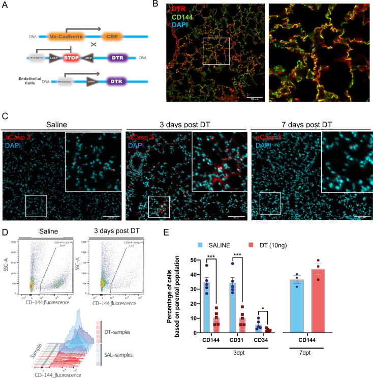
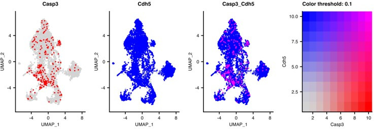
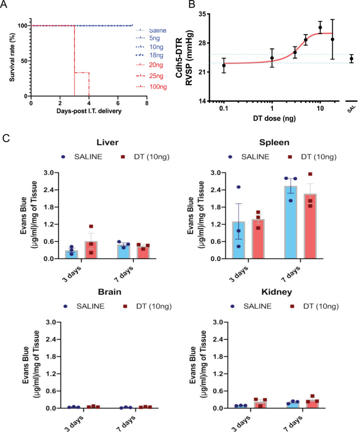
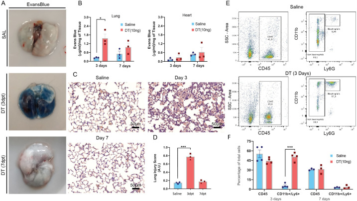
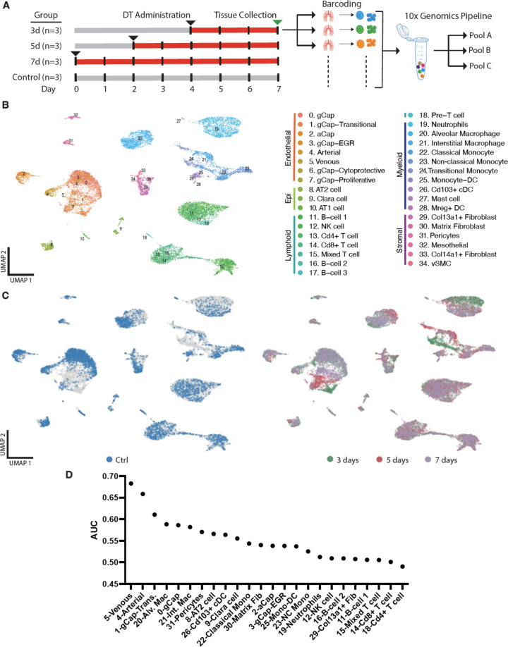
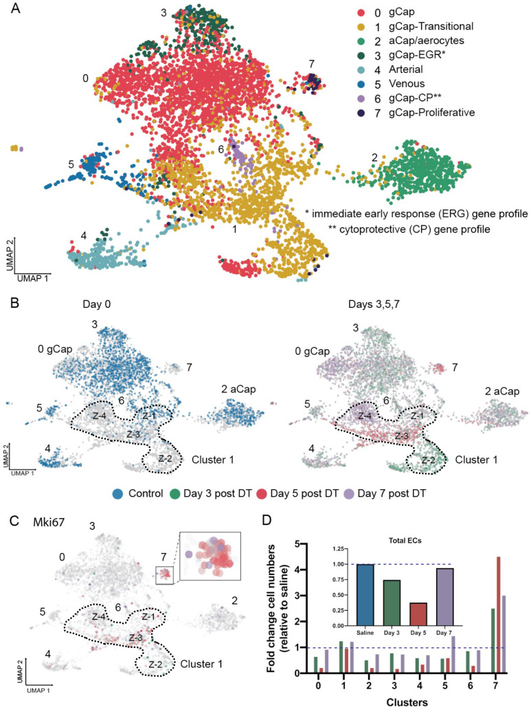
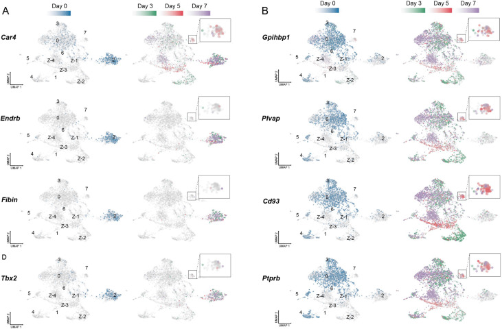
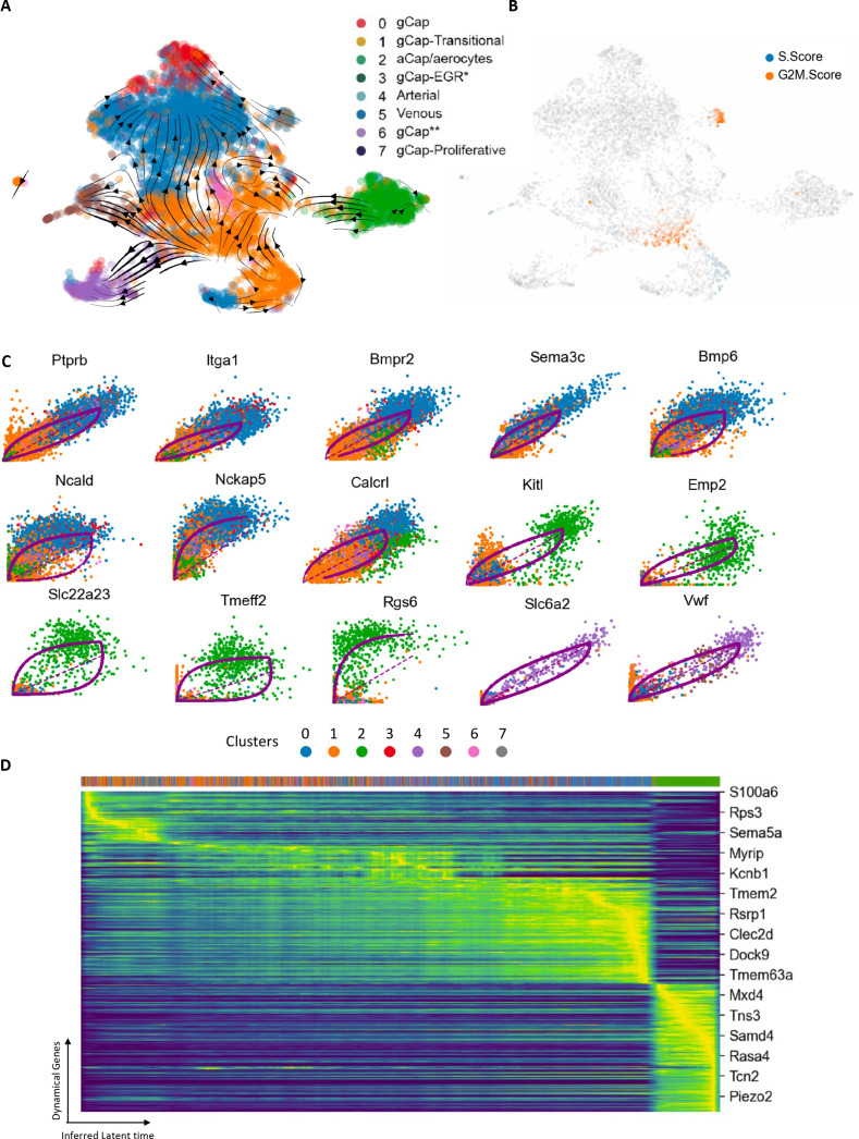
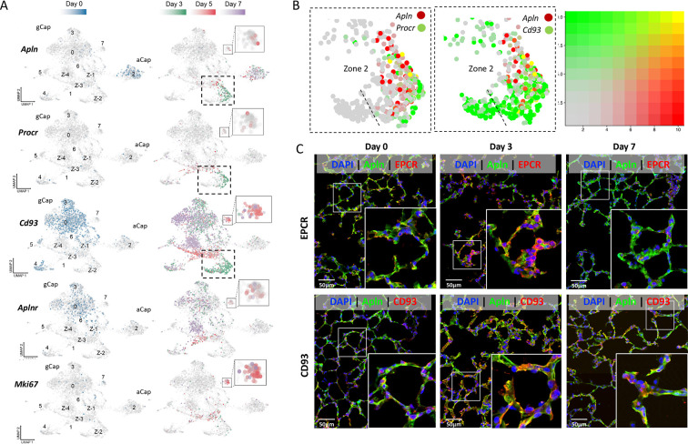
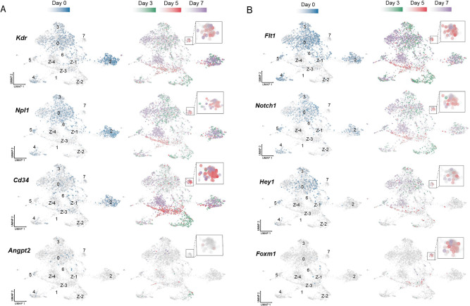
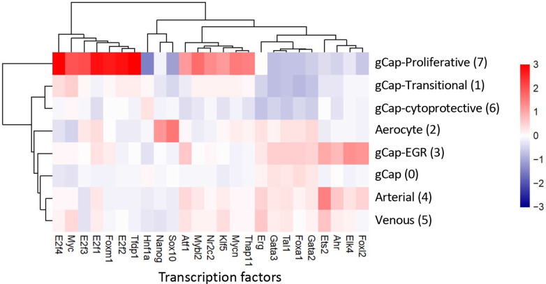
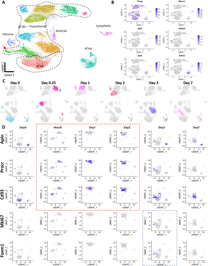
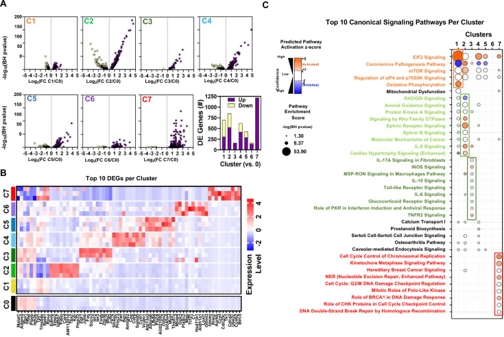
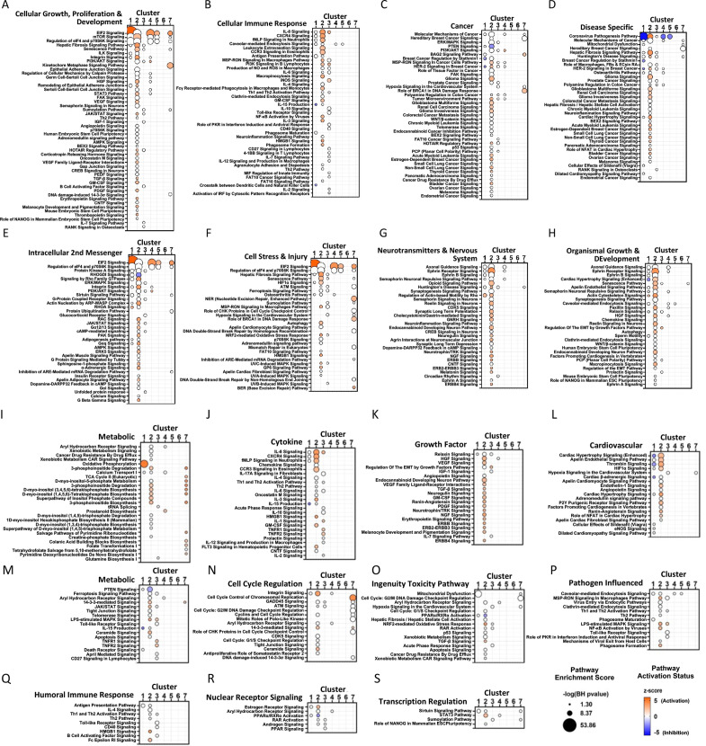
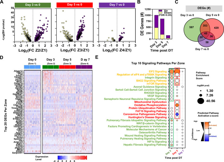
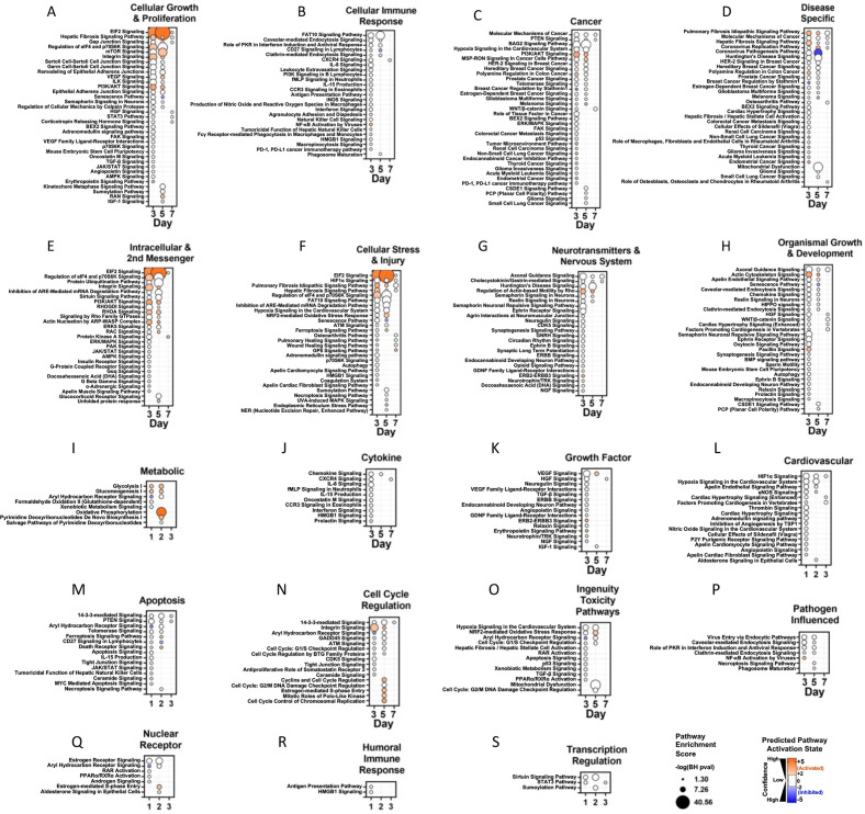
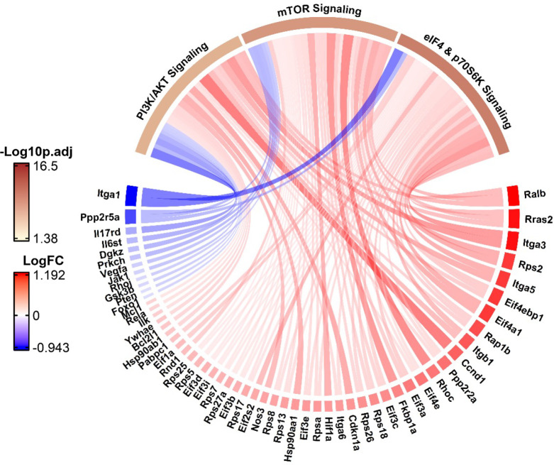
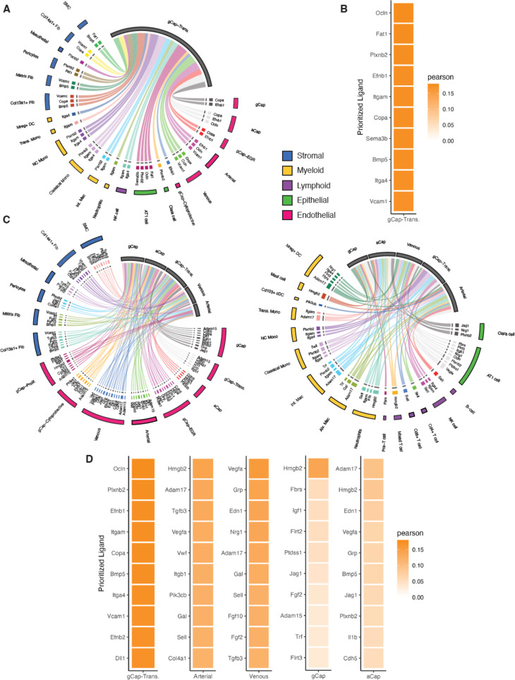
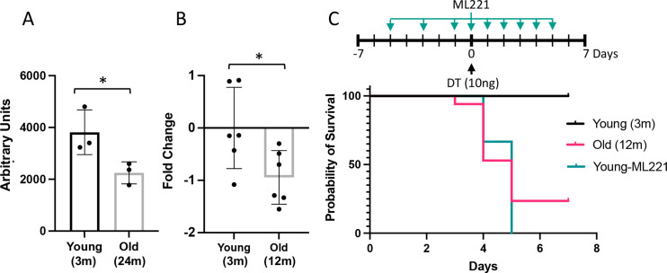
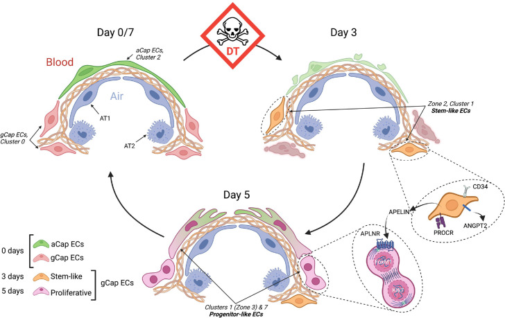
We sought to define the mechanism underlying lung microvascular regeneration in a model of severe acute lung injury (ALI) induced by selective lung endothelial cell ablation. Intratracheal instillation of DT in transgenic mice expressing human diphtheria toxin (DT) receptor targeted to ECs resulted in ablation of >70% of lung ECs, producing severe ALI with near complete resolution by 7 days. Using single-cell RNA sequencing, eight distinct endothelial clusters were resolved, including alveolar aerocytes (aCap) ECs expressing apelin at baseline and general capillary (gCap) ECs expressing the apelin receptor. At 3 days post-injury, a novel gCap EC population emerged characterized by de novo expression of apelin, together with the stem cell marker, protein C receptor. These stem-like cells transitioned at 5 days to proliferative endothelial progenitor-like cells, expressing apelin receptor together with the pro-proliferative transcription factor, , and were responsible for the rapid replenishment of all depleted EC populations by 7 days post-injury. Treatment with an apelin receptor antagonist prevented ALI resolution and resulted in excessive mortality, consistent with a central role for apelin signaling in EC regeneration and microvascular repair. The lung has a remarkable capacity for microvasculature EC regeneration which is orchestrated by newly emergent apelin-expressing gCap endothelial stem-like cells that give rise to highly proliferative, apelin receptor-positive endothelial progenitors responsible for the regeneration of the lung microvasculature.

摘要

我们试图在选择性肺内皮细胞消融诱导的严重急性肺损伤 (ALI) 模型中定义肺微血管再生的机制。在表达靶向内皮细胞的人白喉毒素 (DT) 受体的转基因小鼠中,通过气管内滴注 DT,导致 >70%的肺内皮细胞消融,产生严重的 ALI,在 7 天内几乎完全缓解。使用单细胞 RNA 测序,解析出八个不同的内皮细胞簇,包括在基线时表达阿利克仑的肺泡气细胞 (aCap) 内皮细胞和表达阿利克仑受体的一般毛细血管 (gCap) 内皮细胞。在损伤后 3 天,出现了一种新的 gCap EC 群体,其特征是新表达阿利克仑,同时表达干细胞标记物蛋白 C 受体。这些类干细胞在 5 天过渡到增殖性内皮祖细胞样细胞,表达阿利克仑受体和促增殖转录因子 ,并在损伤后 7 天内迅速补充所有耗尽的 EC 群体。阿利克仑受体拮抗剂治疗可预防 ALI 缓解并导致死亡率过高,这与阿利克仑信号在 EC 再生和微血管修复中的核心作用一致。肺具有非凡的微血管内皮细胞再生能力,由新出现的表达阿利克仑的 gCap 内皮类干细胞协调,这些干细胞产生高度增殖的、阿利克仑受体阳性的内皮祖细胞,负责肺微血管的再生。

https://cdn.ncbi.nlm.nih.gov/pmc/blobs/3873/10181823/5f4555d89d73/elife-80900-sa2-fig1.jpg
https://cdn.ncbi.nlm.nih.gov/pmc/blobs/3873/10181823/c1ecefb2f871/elife-80900-fig1.jpg
https://cdn.ncbi.nlm.nih.gov/pmc/blobs/3873/10181823/43777259026f/elife-80900-fig1-figsupp1.jpg
https://cdn.ncbi.nlm.nih.gov/pmc/blobs/3873/10181823/025737eb24ed/elife-80900-fig2.jpg
https://cdn.ncbi.nlm.nih.gov/pmc/blobs/3873/10181823/b4185ff3aa0c/elife-80900-fig3.jpg
https://cdn.ncbi.nlm.nih.gov/pmc/blobs/3873/10181823/80e4b1f647a3/elife-80900-fig3-figsupp1.jpg
https://cdn.ncbi.nlm.nih.gov/pmc/blobs/3873/10181823/66f90b7bd8b3/elife-80900-fig3-figsupp2.jpg
https://cdn.ncbi.nlm.nih.gov/pmc/blobs/3873/10181823/716d1b378018/elife-80900-fig4.jpg
https://cdn.ncbi.nlm.nih.gov/pmc/blobs/3873/10181823/8195f21cc666/elife-80900-fig4-figsupp1.jpg
https://cdn.ncbi.nlm.nih.gov/pmc/blobs/3873/10181823/89370d43db59/elife-80900-fig5.jpg
https://cdn.ncbi.nlm.nih.gov/pmc/blobs/3873/10181823/64faf721fb20/elife-80900-fig6.jpg
https://cdn.ncbi.nlm.nih.gov/pmc/blobs/3873/10181823/652b9e02bf4e/elife-80900-fig6-figsupp1.jpg
https://cdn.ncbi.nlm.nih.gov/pmc/blobs/3873/10181823/95f3038615ec/elife-80900-fig6-figsupp2.jpg
https://cdn.ncbi.nlm.nih.gov/pmc/blobs/3873/10181823/e38cad4c9128/elife-80900-fig6-figsupp3.jpg
https://cdn.ncbi.nlm.nih.gov/pmc/blobs/3873/10181823/567fe69fcf18/elife-80900-fig7.jpg
https://cdn.ncbi.nlm.nih.gov/pmc/blobs/3873/10181823/72e6c32187df/elife-80900-fig7-figsupp1.jpg
https://cdn.ncbi.nlm.nih.gov/pmc/blobs/3873/10181823/02c9156efa83/elife-80900-fig8.jpg
https://cdn.ncbi.nlm.nih.gov/pmc/blobs/3873/10181823/fc3a97da6e48/elife-80900-fig8-figsupp1.jpg
https://cdn.ncbi.nlm.nih.gov/pmc/blobs/3873/10181823/f45f92912702/elife-80900-fig8-figsupp2.jpg
https://cdn.ncbi.nlm.nih.gov/pmc/blobs/3873/10181823/d98d996d786b/elife-80900-fig9.jpg
https://cdn.ncbi.nlm.nih.gov/pmc/blobs/3873/10181823/dd641a83e392/elife-80900-fig10.jpg
https://cdn.ncbi.nlm.nih.gov/pmc/blobs/3873/10181823/dd5a49982312/elife-80900-fig11.jpg
https://cdn.ncbi.nlm.nih.gov/pmc/blobs/3873/10181823/5f4555d89d73/elife-80900-sa2-fig1.jpg

相似文献

1
Single-cell transcriptomic atlas of lung microvascular regeneration after targeted endothelial cell ablation.靶向内皮细胞消融后肺微血管再生的单细胞转录组图谱。
Elife. 2023 Apr 20;12:e80900. doi: 10.7554/eLife.80900.
2
ABCG2-Expressing Clonal Repopulating Endothelial Cells Serve to Form and Maintain Blood Vessels.ABCG2 表达的克隆性造血内皮细胞有助于血管的形成和维持。
Circulation. 2024 Aug 6;150(6):451-465. doi: 10.1161/CIRCULATIONAHA.122.061833. Epub 2024 Apr 29.
3
Bone morphogenetic protein 10 serves as a biomarker and a potential therapeutic target for endothelial dysfunction in endotoxin-induced acute lung injury.骨形态发生蛋白10作为内毒素诱导的急性肺损伤中内皮功能障碍的生物标志物和潜在治疗靶点。
J Transl Med. 2025 Jul 8;23(1):755. doi: 10.1186/s12967-025-06742-6.
4
Electronic cigarettes for smoking cessation and reduction.用于戒烟和减少吸烟量的电子烟。
Cochrane Database Syst Rev. 2014(12):CD010216. doi: 10.1002/14651858.CD010216.pub2. Epub 2014 Dec 17.
5
HYDROGEN PREVENTS LIPOPOLYSACCHARIDE-INDUCED PULMONARY MICROVASCULAR ENDOTHELIAL CELL INJURY BY INHIBITING STORE-OPERATED Ca 2+ ENTRY REGULATED BY STIM1/ORAI1.氢气通过抑制由基质相互作用分子1/钙释放激活钙通道蛋白1调控的钙库操纵性钙内流来预防脂多糖诱导的肺微血管内皮细胞损伤。
Shock. 2024 May 1;61(5):766-775. doi: 10.1097/SHK.0000000000002279. Epub 2023 Nov 20.
6
Electronic cigarettes for smoking cessation.电子烟戒烟。
Cochrane Database Syst Rev. 2022 Nov 17;11(11):CD010216. doi: 10.1002/14651858.CD010216.pub7.
7
MTOR maintains endothelial cell integrity to limit lung vascular injury.雷帕霉素靶蛋白维持内皮细胞完整性以限制肺血管损伤。
J Biol Chem. 2024 Dec;300(12):107952. doi: 10.1016/j.jbc.2024.107952. Epub 2024 Nov 6.
8
Electronic cigarettes for smoking cessation.电子烟戒烟。
Cochrane Database Syst Rev. 2021 Sep 14;9(9):CD010216. doi: 10.1002/14651858.CD010216.pub6.
9
Mesenchymal stem cell-secreted KGF ameliorates acute lung injury via the Gab1/ERK/NF-κB signaling axis.间充质干细胞分泌的角质形成细胞生长因子通过Gab1/ERK/NF-κB信号轴改善急性肺损伤。
Cell Mol Biol Lett. 2025 Jul 10;30(1):79. doi: 10.1186/s11658-025-00757-z.
10
Endothelial cell-derived extracellular vesicles modulate the therapeutic efficacy of mesenchymal stem cells through IDH2/TET pathway in ARDS.内皮细胞衍生的细胞外囊泡通过 IDH2/TET 通路调节骨髓间充质干细胞在急性呼吸窘迫综合征中的治疗效果。
Cell Commun Signal. 2024 May 27;22(1):293. doi: 10.1186/s12964-024-01672-0.

引用本文的文献

1
APJ regulates the balance between self-renewal and differentiation of vascular endothelial stem cells.APJ调节血管内皮干细胞自我更新与分化之间的平衡。
Inflamm Regen. 2025 Aug 4;45(1):25. doi: 10.1186/s41232-025-00389-y.
2
The Destructive Cycle in Bronchopulmonary Dysplasia: The Rationale for Systems Pharmacology Therapeutics.支气管肺发育不良中的破坏循环:系统药理学治疗原理
Antioxidants (Basel). 2025 Jul 10;14(7):844. doi: 10.3390/antiox14070844.
3
Truncated NTRK2 is induced in CAP1 endothelial cells during mouse lung injury-repair.

本文引用的文献

1
decoupleR: ensemble of computational methods to infer biological activities from omics data.decoupleR:用于从组学数据推断生物活性的计算方法集合。
Bioinform Adv. 2022 Mar 8;2(1):vbac016. doi: 10.1093/bioadv/vbac016. eCollection 2022.
2
COVID-19-associated Lung Microvascular Endotheliopathy: A "From the Bench" Perspective.COVID-19 相关肺微血管内皮病变:从基础研究角度看。
Am J Respir Crit Care Med. 2022 Oct 15;206(8):961-972. doi: 10.1164/rccm.202107-1774OC.
3
Single-cell transcriptomic profiling of lung endothelial cells identifies dynamic inflammatory and regenerative subpopulations.
在小鼠肺损伤修复过程中,截短的NTRK2在CAP1内皮细胞中被诱导产生。
iScience. 2025 Jun 24;28(7):112973. doi: 10.1016/j.isci.2025.112973. eCollection 2025 Jul 18.
4
Unraveling the deadly dance: endothelial cells and neutrophils in sepsis-induced acute lung injury/acute respiratory distress syndrome.解析致命之舞:脓毒症诱导的急性肺损伤/急性呼吸窘迫综合征中的内皮细胞与中性粒细胞
Front Cell Dev Biol. 2025 May 22;13:1551138. doi: 10.3389/fcell.2025.1551138. eCollection 2025.
5
Human endothelial colony forming cells (ECFCs) require endothelial protein C receptor (EPCR) for cell cycle progression and angiogenic activity.人内皮集落形成细胞(ECFCs)的细胞周期进程和血管生成活性需要内皮蛋白C受体(EPCR)。
Angiogenesis. 2025 May 23;28(3):30. doi: 10.1007/s10456-025-09982-8.
6
Single-cell transcriptional responses of T cells during microsporidia infection.微孢子虫感染期间T细胞的单细胞转录反应。
Commun Biol. 2025 Apr 5;8(1):567. doi: 10.1038/s42003-025-07990-4.
7
PRMT1 Ablation in Endothelial Cells Causes Endothelial Dysfunction and Aggravates COPD Attributable to Dysregulated NF-κB Signaling.内皮细胞中PRMT1缺失导致内皮功能障碍,并因NF-κB信号失调而加重慢性阻塞性肺疾病。
Adv Sci (Weinh). 2025 May;12(19):e2411514. doi: 10.1002/advs.202411514. Epub 2025 Mar 26.
8
Unveiling the immunomodulatory dance: endothelial cells' function and their role in non-small cell lung cancer.揭示免疫调节之舞:内皮细胞的功能及其在非小细胞肺癌中的作用。
Mol Cancer. 2025 Jan 16;24(1):21. doi: 10.1186/s12943-024-02221-6.
9
Polyclonal regeneration of mouse bone marrow endothelial cells after irradiative conditioning.辐照预处理后小鼠骨髓内皮细胞的多克隆再生。
Cell Rep. 2024 Nov 26;43(11):114779. doi: 10.1016/j.celrep.2024.114779. Epub 2024 Oct 26.
10
Process-Specific Blood Biomarkers and Outcomes in COVID-19 Versus Non-COVID-19 ARDS (APEL-COVID Study): A Prospective, Observational Cohort Study.新型冠状病毒肺炎与非新型冠状病毒肺炎急性呼吸窘迫综合征的特定病程血液生物标志物及预后(APEL-COVID研究):一项前瞻性观察性队列研究
J Clin Med. 2024 Oct 4;13(19):5919. doi: 10.3390/jcm13195919.
单细胞转录组谱分析鉴定出肺血管内皮细胞中具有动态炎症和再生特征的亚群。
JCI Insight. 2022 Jun 8;7(11):e158079. doi: 10.1172/jci.insight.158079.
4
CellRank for directed single-cell fate mapping.细胞排序用于有向单细胞命运图谱绘制。
Nat Methods. 2022 Feb;19(2):159-170. doi: 10.1038/s41592-021-01346-6. Epub 2022 Jan 13.
5
Integrated Single-Cell Atlas of Endothelial Cells of the Human Lung.人类肺部血管内皮细胞的综合单细胞图谱
Circulation. 2021 Jul 27;144(4):286-302. doi: 10.1161/CIRCULATIONAHA.120.052318. Epub 2021 May 25.
6
Endothelial activation and dysfunction in COVID-19: from basic mechanisms to potential therapeutic approaches.新型冠状病毒肺炎中的内皮细胞激活与功能障碍:从基本机制到潜在治疗方法
Signal Transduct Target Ther. 2020 Dec 24;5(1):293. doi: 10.1038/s41392-020-00454-7.
7
muscat detects subpopulation-specific state transitions from multi-sample multi-condition single-cell transcriptomics data.Muscat 可从多样本多条件单细胞转录组学数据中检测到亚群特异性状态转变。
Nat Commun. 2020 Nov 30;11(1):6077. doi: 10.1038/s41467-020-19894-4.
8
Pulmonary pathology of ARDS in COVID-19: A pathological review for clinicians.新型冠状病毒肺炎急性呼吸窘迫综合征的肺部病理学:临床医生的病理评估。
Respir Med. 2021 Jan;176:106239. doi: 10.1016/j.rmed.2020.106239. Epub 2020 Nov 19.
9
Capillary cell-type specialization in the alveolus.肺泡毛细血管细胞类型特化。
Nature. 2020 Oct;586(7831):785-789. doi: 10.1038/s41586-020-2822-7. Epub 2020 Oct 14.
10
Severe COVID-19 Is a Microvascular Disease.重症新型冠状病毒肺炎是一种微血管疾病。
Circulation. 2020 Oct 27;142(17):1609-1611. doi: 10.1161/CIRCULATIONAHA.120.050354. Epub 2020 Sep 2.