Department of Internal Medicine (463), Radboud University Medical Center, PO Box 9101, Geert Grooteplein 8, Nijmegen 6500 HB, The Netherlands.
Division of Human Nutrition and Health, Wageningen University and Research Division of Human Nutrition and Health (Bode 62), P.O. Box 176700 AA, Wageningen, The Netherlands.
Cardiovasc Res. 2023 Aug 19;119(10):1942-1951. doi: 10.1093/cvr/cvad058.
The article investigates whether chronic hyperglycaemia in Type 1 diabetes (T1D) is associated with a proinflammatory immune signature and with arterial wall inflammation, driving the development of atherosclerosis.
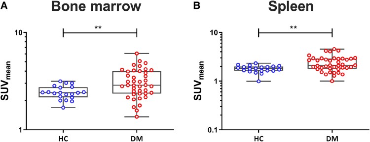
Patients with T1D (n = 41), and healthy age-, sex-, and body mass index-matched controls (n = 20) were recruited. Arterial wall inflammation and haematopoietic activity were measured with 2'-deoxy-2'-(18F)-fluoro-D-glucose (18F-FDG) positron emission tomography/computed tomography. In addition, flow cytometry of circulating leucocytes was performed as well as targeted proteomics to measure circulating inflammatory markers. 18F-FDG uptake in the wall of the abdominal aorta, carotid arteries, and iliac arteries was higher in T1D compared with that in the healthy controls. Also, 18F-FDG uptake in the bone marrow and spleen was higher in patients with T1D. CCR2 and CD36 expressions on circulating monocytes were higher in patients with T1D, as well as several circulating inflammatory proteins. In addition, several circulating inflammatory markers (osteoprotegerin, transforming growth factor-alpha, CX3CL1, and colony-stimulating factor-1) displayed a positive correlation with FDG uptake. Within T1D, no differences were found between people with a high and low HbA1c.
These findings strengthen the concept that chronic hyperglycaemia in T1D induces inflammatory changes that fuel arterial wall inflammation leading to atherosclerosis. The degree of hyperglycaemia appears to play a minor role in driving this inflammatory response in patients with T1D.
本文旨在研究 1 型糖尿病(T1D)患者的慢性高血糖是否与促炎免疫特征以及动脉壁炎症有关,进而导致动脉粥样硬化的发生。
研究纳入了 41 名 T1D 患者和 20 名年龄、性别和体重指数匹配的健康对照者。使用 2'-脱氧-2'-(18F)-氟-D-葡萄糖(18F-FDG)正电子发射断层扫描/计算机断层扫描来测量动脉壁炎症和造血活性。此外,还进行了循环白细胞的流式细胞术以及靶向蛋白质组学来测量循环炎症标志物。与健康对照组相比,T1D 患者的腹主动脉、颈动脉和髂动脉壁 18F-FDG 摄取更高。此外,T1D 患者的骨髓和脾脏中 18F-FDG 摄取也更高。循环单核细胞上的 CCR2 和 CD36 表达在 T1D 患者中更高,同时还有几种循环炎症蛋白。此外,几种循环炎症标志物(骨保护素、转化生长因子-α、CX3CL1 和集落刺激因子-1)与 FDG 摄取呈正相关。在 T1D 患者中,HbA1c 水平高和低的患者之间没有差异。
这些发现进一步证实了慢性高血糖会引起 T1D 患者的炎症变化,从而导致动脉壁炎症和动脉粥样硬化的发生。高血糖的程度似乎在驱动 T1D 患者的这种炎症反应中作用较小。