Robotics and Mechatronics, University of Twente, Drienerlolaan 5, 7522 NB, Enschede, The Netherlands.
Department of Urology, Ziekenhuisgroep Twente (ZGT), Zilvermeeuw 1, 7609 PP, Almelo, The Netherlands.
Int J Comput Assist Radiol Surg. 2023 Oct;18(10):1915-1924. doi: 10.1007/s11548-023-02900-7. Epub 2023 Apr 21.
Abnormalities in the bladder wall require careful investigation regarding type, spatial position and invasiveness. Construction of a 3-D model of the bladder is helpful to ensure adequate coverage of the scanning procedure, quantitative comparison of bladder wall textures between successive sessions and finding back previously discovered abnormalities.
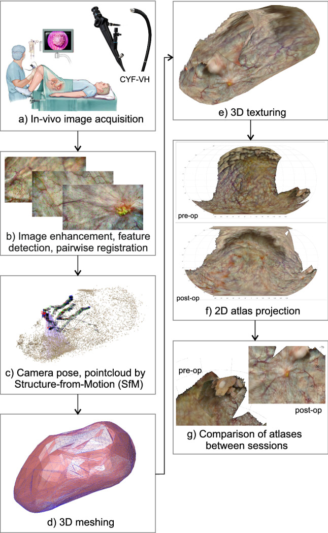
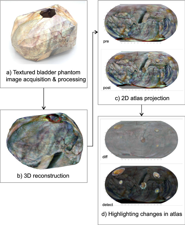
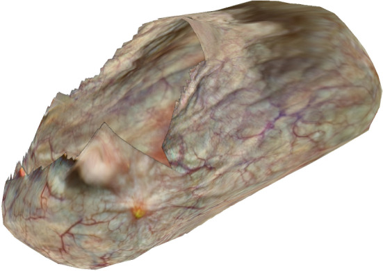
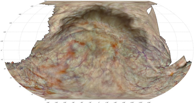
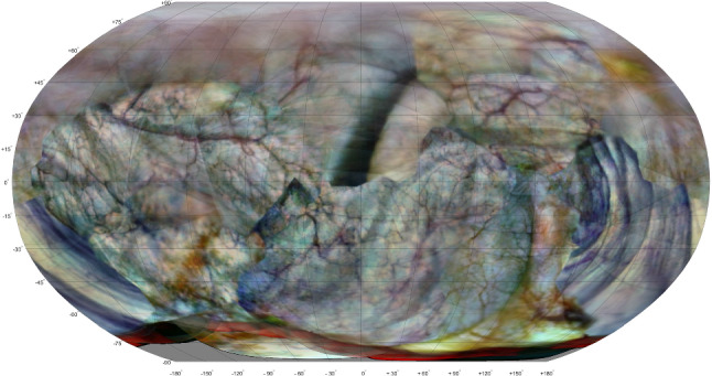
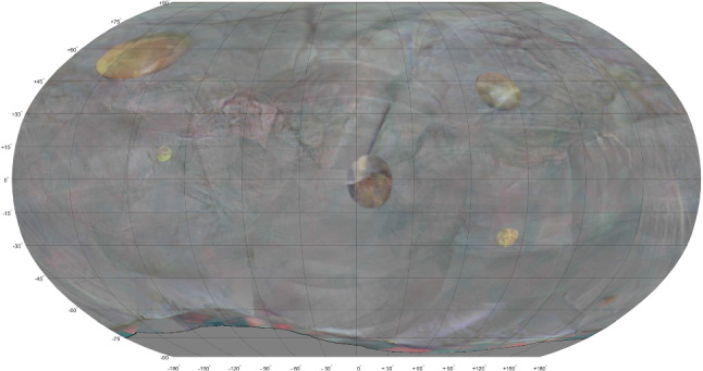
Videos of both an in vivo bladder and a textured bladder phantom were acquired. Structure-from-motion and bundle adjustment algorithms were used to construct a 3-D point cloud, approximate it by a surface mesh, texture it with the back-projected camera frames and draw the corresponding 2-D atlas. Reconstructions of successive sessions were compared; those of the bladder phantom were co-registered, transformed using 3-D thin plate splines and post-processed to highlight significant changes in texture.
The reconstruction algorithms of the presented workflow were able to construct 3-D models and corresponding 2-D atlas of both the in vivo bladder and the bladder phantom. For the in vivo bladder the portion of the reconstructed surface area was 58% and 79% for the pre- and post-operative scan, respectively. For the bladder phantom the full surface was reconstructed and the mean reprojection error was 0.081 mm (range 0-0.79 mm). In inter-session comparison the changes in texture were correctly indicated for all six locations.
The proposed proof of concept was able to perform 3-D and 2-D reconstruction of an in vivo bladder wall based on a set of monocular images. In a phantom study the computer vision algorithms were also effective in co-registering reconstructions of successive sessions and highlighting texture changes between sessions. These techniques may be useful for detecting, monitoring and revisiting suspicious lesions.
膀胱壁的异常需要仔细检查其类型、空间位置和侵袭性。构建膀胱的 3D 模型有助于确保扫描过程得到充分覆盖,对连续阶段的膀胱壁纹理进行定量比较,并找到先前发现的异常。
获取活体膀胱和纹理膀胱模型的视频。使用运动结构和捆绑调整算法构建 3D 点云,用曲面网格逼近它,用反向投影的相机帧对其进行纹理处理,并绘制相应的 2D 图谱。比较连续阶段的重建;对膀胱模型进行配准,使用 3D 薄板样条进行变换,并进行后处理以突出纹理的显著变化。
所提出工作流程的重建算法能够构建活体膀胱和膀胱模型的 3D 模型和相应的 2D 图谱。对于活体膀胱,重建表面面积的部分分别为术前和术后扫描的 58%和 79%。对于膀胱模型,完全重建了表面,平均重投影误差为 0.081 毫米(范围为 0-0.79 毫米)。在阶段间比较中,所有六个位置的纹理变化都被正确指示。
该概念验证能够基于一组单目图像对活体膀胱壁进行 3D 和 2D 重建。在模型研究中,计算机视觉算法还能够有效地对连续阶段的重建进行配准,并突出阶段间的纹理变化。这些技术可能有助于检测、监测和复查可疑病变。