• 文献检索
  • 文档翻译
  • 深度研究
  • 学术资讯
  • Suppr Zotero 插件Zotero 插件
  • 邀请有礼
  • 套餐&价格
  • 历史记录
应用&插件
Suppr Zotero 插件Zotero 插件浏览器插件Mac 客户端Windows 客户端微信小程序
定价
高级版会员购买积分包购买API积分包
服务
文献检索文档翻译深度研究API 文档MCP 服务
关于我们
关于 Suppr公司介绍联系我们用户协议隐私条款
关注我们

Suppr 超能文献

核心技术专利:CN118964589B侵权必究
粤ICP备2023148730 号-1Suppr @ 2026

文献检索

告别复杂PubMed语法,用中文像聊天一样搜索,搜遍4000万医学文献。AI智能推荐,让科研检索更轻松。

立即免费搜索

文件翻译

保留排版,准确专业,支持PDF/Word/PPT等文件格式,支持 12+语言互译。

免费翻译文档

深度研究

AI帮你快速写综述,25分钟生成高质量综述,智能提取关键信息,辅助科研写作。

立即免费体验

pT1b 期食管鳞癌中淋巴管脉管侵犯的预后意义。

Prognostic significance of lymphovascular invasion in patients with pT1b esophageal squamous cell carcinoma.

机构信息

Department of Pathology, National Cancer Center/National Clinical Research Center for Cancer/Cancer Hospital, Chinese Academy of Medical Sciences and Peking Union Medical College, Beijing, China.

Department of Medical Record, National Cancer Center/National Clinical Research Center for Cancer/Cancer Hospital, Chinese Academy of Medical Sciences and Peking Union Medical College, Beijing, 100021, China.

出版信息

BMC Cancer. 2023 Apr 22;23(1):370. doi: 10.1186/s12885-023-10858-7.

DOI:10.1186/s12885-023-10858-7
PMID:37087442
原文链接:https://pmc.ncbi.nlm.nih.gov/articles/PMC10122816/
Abstract

BACKGROUND

Lymphovascular invasion (LVI) is a crucial predictor of lymph node metastasis (LNM). However, few studies have investigated the LVI positivity rate and its clinical significance in pT1b esophageal squamous cell carcinoma (ESCC) using immunohistochemistry and elastin staining.

METHODS

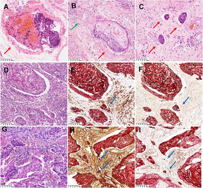
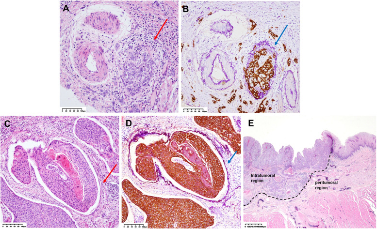
We collected data from158 patients with pT1b ESCC who had undergone radical esophagectomy. All paraffin blocks of invasive carcinoma from each patient were subjected to HE staining, elastin staining + CK (AE1/AE3) immunohistochemistry (E&IHC), and CD31/D2-40 + CK (AE1/AE3) double immunohistochemistry (D-IHC). The LVI was classified into types, i.e., vascular invasion (VI) and lymphatic vessel invasion (LI), and its location, quantity, and clinical significance were explored.

RESULTS

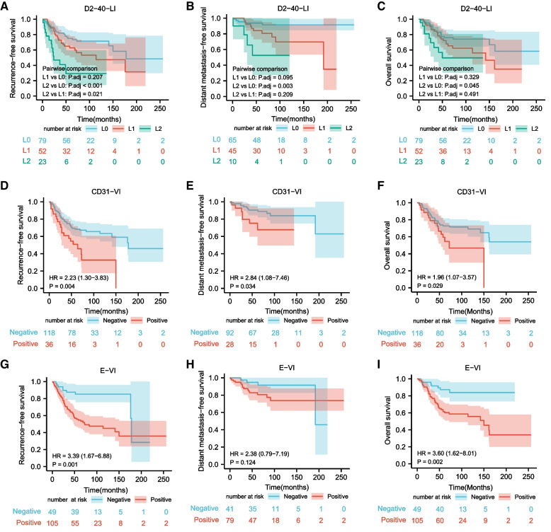
The positivity rates of VI by E&IHC (E-VI), VI by CD31D-IHC (CD31-VI), and LI by D2-40 D-IHC (D2-40-LI) were significantly higher than those obtained by HE staining (P < 0.001, respectively). CD31-VI and E-VI were independent adverse prognostic factors for recurrence-free survival (RFS), and they were significantly associated with poor distant metastasis-free survival and overall survival in pT1b ESCC. Intratumoral LVI was also crucial in pT1b ESCC, and L2 (the count of D2-40-LI was 5 or more) was the strongest predictor for LNM and RFS in pT1b ESCC.

CONCLUSION

E&IHC and D-IHC can dramatically improve the detection rate of LVI in pT1b ESCC, and the classification and grading of LVI can help to improve the prediction of LNM and prognosis.

摘要

背景

淋巴管浸润(LVI)是淋巴结转移(LNM)的重要预测因子。然而,使用免疫组织化学和弹力纤维染色,很少有研究调查 pT1b 食管鳞状细胞癌(ESCC)中的 LVI 阳性率及其临床意义。

方法

我们收集了 158 例接受根治性食管切除术的 pT1b ESCC 患者的数据。每位患者的浸润性癌石蜡块均行 HE 染色、弹力纤维染色+CK(AE1/AE3)免疫组化(E&IHC)和 CD31/D2-40+CK(AE1/AE3)双重免疫组化(D-IHC)。LVI 分为血管浸润(VI)和淋巴管浸润(LI)两种类型,探讨其位置、数量及临床意义。

结果

E&IHC(E-VI)、CD31 D-IHC(CD31-VI)和 D2-40 D-IHC(D2-40-LI)的 VI 阳性率均显著高于 HE 染色(P<0.001)。CD31-VI 和 E-VI 是无复发生存(RFS)的独立不良预后因素,与 pT1b ESCC 患者的远处无转移生存和总生存不良显著相关。肿瘤内 LVI 在 pT1b ESCC 中也很重要,L2(D2-40-LI 计数为 5 个或更多)是预测 pT1b ESCC 中 LNM 和 RFS 的最强预测因子。

结论

E&IHC 和 D-IHC 可显著提高 pT1b ESCC 中 LVI 的检出率,LVI 的分类和分级有助于提高 LNM 和预后的预测。

https://cdn.ncbi.nlm.nih.gov/pmc/blobs/f7a3/10122816/4414be26a67b/12885_2023_10858_Fig3_HTML.jpg
https://cdn.ncbi.nlm.nih.gov/pmc/blobs/f7a3/10122816/66311a22fff2/12885_2023_10858_Fig1_HTML.jpg
https://cdn.ncbi.nlm.nih.gov/pmc/blobs/f7a3/10122816/a09735b6eb4a/12885_2023_10858_Fig2_HTML.jpg
https://cdn.ncbi.nlm.nih.gov/pmc/blobs/f7a3/10122816/4414be26a67b/12885_2023_10858_Fig3_HTML.jpg
https://cdn.ncbi.nlm.nih.gov/pmc/blobs/f7a3/10122816/66311a22fff2/12885_2023_10858_Fig1_HTML.jpg
https://cdn.ncbi.nlm.nih.gov/pmc/blobs/f7a3/10122816/a09735b6eb4a/12885_2023_10858_Fig2_HTML.jpg
https://cdn.ncbi.nlm.nih.gov/pmc/blobs/f7a3/10122816/4414be26a67b/12885_2023_10858_Fig3_HTML.jpg

相似文献

1
Prognostic significance of lymphovascular invasion in patients with pT1b esophageal squamous cell carcinoma.pT1b 期食管鳞癌中淋巴管脉管侵犯的预后意义。
BMC Cancer. 2023 Apr 22;23(1):370. doi: 10.1186/s12885-023-10858-7.
2
Tumor budding and tumor-infiltrating lymphocytes can predict prognosis in pT1b esophageal squamous cell carcinoma.肿瘤芽和肿瘤浸润淋巴细胞可预测 pT1b 食管鳞癌的预后。
Thorac Cancer. 2023 Sep;14(25):2608-2617. doi: 10.1111/1759-7714.15043. Epub 2023 Jul 19.
3
The role of vascular invasion and lymphatic invasion in predicting recurrent thoracic oesophageal squamous cell carcinoma.血管侵犯和淋巴管侵犯在预测复发性胸段食管鳞癌中的作用。
World J Surg Oncol. 2022 Jan 10;20(1):12. doi: 10.1186/s12957-021-02458-1.
4
Lymphatic vessel invasion detected by the D2-40 monoclonal antibody is an independent prognostic factor in node-negative esophageal squamous cell carcinoma.D2-40 单克隆抗体检测到的淋巴管侵犯是淋巴结阴性食管鳞癌的独立预后因素。
J Surg Oncol. 2012 Mar;105(3):277-83. doi: 10.1002/jso.22079. Epub 2011 Aug 24.
5
Clinicopathological investigation of lymph node metastasis predictors in superficial esophageal squamous cell carcinoma with a focus on evaluation of lympho-vascular invasion.以评估淋巴管侵犯为重点的食管浅表鳞状细胞癌淋巴结转移预测因素的临床病理研究
Scand J Gastroenterol. 2013 Oct;48(10):1173-82. doi: 10.3109/00365521.2013.832365. Epub 2013 Aug 26.
6
Identification and Validation of Lymphovascular Invasion as a Prognostic and Staging Factor in Node-Negative Esophageal Squamous Cell Carcinoma.淋巴结阴性食管鳞癌中淋巴管浸润的鉴定和验证及其作为预后和分期因素的意义。
J Thorac Oncol. 2016 Apr;11(4):583-92. doi: 10.1016/j.jtho.2015.12.109. Epub 2016 Jan 11.
7
Lymphovascular invasion as an independent prognostic indicator in radically resected thoracic esophageal squamous cell carcinoma.脉管侵犯是根治性切除的胸段食管鳞癌的独立预后指标。
Thorac Cancer. 2019 Feb;10(2):150-155. doi: 10.1111/1759-7714.12922. Epub 2018 Nov 28.
8
Prognostic significance of lymphovascular invasion in patients with esophageal squamous cell carcinoma treated with neoadjuvant chemoradiotherapy.新辅助放化疗治疗的食管鳞状细胞癌患者中淋巴管侵犯的预后意义
Ann Surg Oncol. 2015 Jan;22(1):338-43. doi: 10.1245/s10434-014-3881-5. Epub 2014 Jul 15.
9
Intratumoral and peritumoral lymphovascular invasion detected by D2-40 immunohistochemistry correlates with metastasis in primary cutaneous Merkel cell carcinoma.D2-40 免疫组织化学检测的肿瘤内和肿瘤周围淋巴管血管侵犯与原发性皮肤 Merkel 细胞癌的转移相关。
Hum Pathol. 2018 Jul;77:98-107. doi: 10.1016/j.humpath.2018.03.017. Epub 2018 Mar 28.
10
Tumor cell invasion in blood vessels assessed by immunohistochemistry is related to decreased survival in patients with bladder cancer treated with radical cystectomy.免疫组织化学评估的肿瘤细胞血管侵犯与接受根治性膀胱切除术治疗的膀胱癌患者的生存降低有关。
Diagn Pathol. 2021 Nov 22;16(1):109. doi: 10.1186/s13000-021-01171-7.

引用本文的文献

1
Computed tomography-based radiomics nomogram for prediction of lympho-vascular and perineural invasion in esophageal squamous cell cancer patients: a retrospective cohort study.基于计算机断层扫描的放射组学列线图预测食管鳞癌患者的淋巴血管和神经周围侵犯:一项回顾性队列研究。
Cancer Imaging. 2024 Oct 4;24(1):131. doi: 10.1186/s40644-024-00781-w.
2
Quality control indices for standardized diagnosis and treatment of esophageal cancer in China (2022 edition).中国食管癌标准化诊疗质量控制指标(2022年版)
J Natl Cancer Cent. 2023 Aug 11;3(3):167-174. doi: 10.1016/j.jncc.2023.07.005. eCollection 2023 Sep.
3
The Usefulness of Elastin Staining to Detect Vascular Invasion in Cancer.

本文引用的文献

1
Tumor cell invasion in blood vessels assessed by immunohistochemistry is related to decreased survival in patients with bladder cancer treated with radical cystectomy.免疫组织化学评估的肿瘤细胞血管侵犯与接受根治性膀胱切除术治疗的膀胱癌患者的生存降低有关。
Diagn Pathol. 2021 Nov 22;16(1):109. doi: 10.1186/s13000-021-01171-7.
2
Prediction of lymph node metastasis in superficial esophageal squamous cell carcinoma in Asia: a systematic review and meta-analysis.亚洲表浅性食管鳞癌淋巴结转移预测:系统回顾和荟萃分析。
Dis Esophagus. 2020 Dec 7;33(12). doi: 10.1093/dote/doaa032.
3
Clinical features of metastasis from superficial squamous cell carcinoma of the thoracic esophagus.
弹性纤维染色在检测癌症血管侵犯中的作用。
Int J Mol Sci. 2023 Oct 17;24(20):15264. doi: 10.3390/ijms242015264.
胸段食管浅表鳞癌转移的临床特征。
Surgery. 2019 Dec;166(6):1033-1040. doi: 10.1016/j.surg.2019.07.022. Epub 2019 Sep 5.
4
Risk of Lymph Node Metastasis in Patients with the Superficial Spreading Type of Esophageal Squamous Cell Carcinoma.浅表扩散型食管鳞癌患者的淋巴结转移风险。
Digestion. 2020;101(3):239-244. doi: 10.1159/000499017. Epub 2019 Mar 25.
5
Prognostic impact of lymphovascular invasion in lymph node-negative superficial esophageal squamous cell carcinoma.淋巴结阴性的食管浅表鳞癌中淋巴管浸润的预后影响。
Dis Esophagus. 2019 Dec 30;32(11). doi: 10.1093/dote/doz001.
6
Clinicopathological parameters predicting recurrence of pT1N0 esophageal squamous cell carcinoma.预测 pT1N0 食管鳞癌复发的临床病理参数。
World J Gastroenterol. 2018 Dec 7;24(45):5154-5166. doi: 10.3748/wjg.v24.i45.5154.
7
Lymphovascular invasion as an independent prognostic indicator in radically resected thoracic esophageal squamous cell carcinoma.脉管侵犯是根治性切除的胸段食管鳞癌的独立预后指标。
Thorac Cancer. 2019 Feb;10(2):150-155. doi: 10.1111/1759-7714.12922. Epub 2018 Nov 28.
8
A nomogram for predicting lymph node metastasis in surgically resected T1 esophageal squamous cell carcinoma.预测手术切除的T1期食管鳞状细胞癌淋巴结转移的列线图。
J Thorac Dis. 2018 Jul;10(7):4178-4185. doi: 10.21037/jtd.2018.06.51.
9
Predicting lymph node metastasis for endoscopic resection of superficial esophageal squamous cell carcinoma.预测内镜切除浅表性食管鳞状细胞癌的淋巴结转移。
J Thorac Cardiovasc Surg. 2019 Jan;157(1):397-402.e1. doi: 10.1016/j.jtcvs.2018.07.034. Epub 2018 Jul 29.
10
A prediction model for lymph node metastasis in T1 esophageal squamous cell carcinoma.T1 期食管鳞癌淋巴结转移预测模型。
J Thorac Cardiovasc Surg. 2018 Apr;155(4):1902-1908. doi: 10.1016/j.jtcvs.2017.11.005. Epub 2017 Nov 11.