Department of Oncology, The Sidney Kimmel Comprehensive Cancer Center, The Johns Hopkins Hospital, Baltimore, Maryland, USA.
Center for Personalized Cancer Therapy and Division of Hematology and Oncology, Department of Medicine, UC San Diego Moores Cancer Center, La Jolla, California, USA.
Cancer Med. 2023 Jun;12(12):13155-13166. doi: 10.1002/cam4.6000. Epub 2023 May 3.
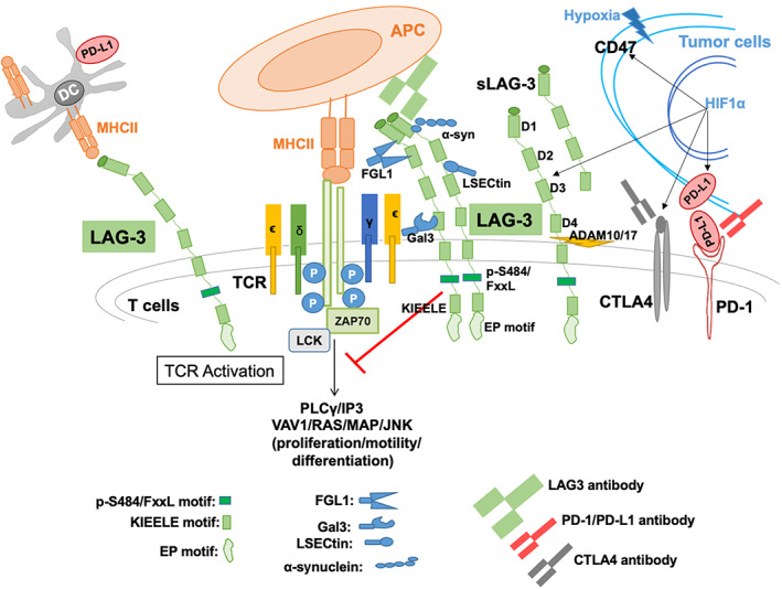
Lymphocyte activation gene 3 (LAG-3) or CD223 is a transmembrane protein that serves as an immune checkpoint which attenuates T-cell activation. Many clinical trials of LAG-3 inhibitors have had modest effects, but recent data indicate that the LAG-3 antibody relatlimab, together with nivolumab (anti-PD-1), provided greater benefit than nivolumab alone in patients with melanoma.
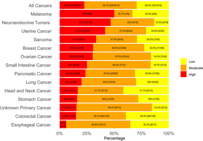
In this study, the RNA expression levels of 397 genes were assessed in 514 diverse cancers at a clinical-grade laboratory (OmniSeq: https://www.omniseq.com/). Transcript abundance was normalized to internal housekeeping gene profiles and ranked (0-100 percentile) using a reference population (735 tumors; 35 histologies).
A total of 116 of 514 tumors (22.6%) had high LAG-3 transcript expression (≥75 percentile rank). Cancers with the greatest proportion of high LAG-3 transcripts were neuroendocrine (47% of patients) and uterine (42%); colorectal had among the lowest proportion of high LAG-3 expression (15% of patients) (all p < 0.05 multivariate); 50% of melanomas were high LAG-3 expressors. There was significant independent association between high LAG-3 expression and high expression of other checkpoints, including programmed death-ligand 1 (PD-L1), PD-1, and CTLA-4, as well as high tumor mutational burden (TMB) ≥10 mutations/megabase, a marker for immunotherapy response (all p < 0.05 multivariate). However, within all tumor types, there was inter-patient variability in LAG-3 expression level.
Prospective studies are therefore needed to determine if high levels of the LAG-3 checkpoint are responsible for resistance to anti-PD-1/PD-L1 or anti-CTLA-4 antibodies. Furthermore, a precision/personalized immunotherapy approach may require interrogating individual tumor immunograms to match patients to the right combination of immunotherapeutic agents for their malignancy.
淋巴细胞激活基因 3(LAG-3)或 CD223 是一种跨膜蛋白,作为免疫检查点,可减弱 T 细胞的激活。许多 LAG-3 抑制剂的临床试验效果都较为温和,但最近的数据表明,LAG-3 抗体 relatlimab 与 nivolumab(抗 PD-1)联合使用,为黑色素瘤患者带来的益处大于 nivolumab 单药治疗。
在这项研究中,在临床级别的实验室(OmniSeq:https://www.omniseq.com/)中评估了 514 种不同癌症中的 397 个基因的 RNA 表达水平。使用参考人群(735 个肿瘤;35 种组织学)对转录本丰度进行标准化并进行排名(0-100 百分位)。
在 514 个肿瘤中,共有 116 个(22.6%)肿瘤的 LAG-3 转录本表达较高(≥75 百分位)。LAG-3 转录本表达较高的癌症包括神经内分泌癌(47%的患者)和子宫癌(42%);结直肠癌患者中 LAG-3 表达较高的比例最低(15%的患者)(所有多变量 p<0.05);50%的黑色素瘤患者为 LAG-3 高表达者。LAG-3 表达较高与其他检查点(包括程序性死亡配体 1(PD-L1)、PD-1 和 CTLA-4)以及高肿瘤突变负荷(TMB)≥10 个突变/兆碱基,这是免疫治疗反应的标志物,都有显著的独立关联(所有多变量 p<0.05)。然而,在所有肿瘤类型中,LAG-3 表达水平存在患者间的差异。
因此,需要进行前瞻性研究以确定 LAG-3 检查点的高水平是否导致对抗 PD-1/PD-L1 或抗 CTLA-4 抗体的耐药性。此外,精确/个性化的免疫治疗方法可能需要检查单个肿瘤免疫组图,以将患者与适合其恶性肿瘤的免疫治疗药物组合相匹配。