Radiology Department, Imam Abdulrahman Bin Faisal University, College of Medicine, Dammam, Saudi Arabia.
Imam Abdulrahman Bin Faisal University, College of Medicine, Dammam, Saudi Arabia.
J Med Life. 2023 Mar;16(3):458-462. doi: 10.25122/jml-2022-0265.
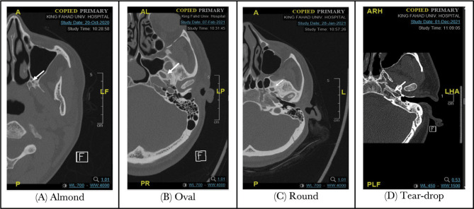
The foramen ovale is one of the essential foramina in the middle cranial fossa, more precisely, in the superior surface of the greater wing of the sphenoid bone. It has essential surgical and diagnostic significance since it serves as a surgical landmark, and crucial neurovascular vessels such as the mandibular nerve and accessory meningeal artery pass through it. Therefore, understanding the morphological and morphometric variations of the foramen ovale is essential for accurately identifying, diagnosing, and managing related pathologies. The study aimed to evaluate the morphological variations and morphometric details of the foramen ovale in the Saudi population. A radiological study was conducted to observe the measurements and the shapes of the foramen ovale in the skull with its anatomical variants. Retrospective data was collected from the Department of Radiology, King Fahad University Hospital, Saudi Arabia. The sample consisted of 100 human skulls from computed tomography scans, including 50 males and 50 females. The values for the mean length, width, and distance from the midline on the right side were 6.462 mm ± 1.681 mm, 4.897 ± 1.0631 mm, and 2.4565 ± 0.51275 mm, and 6.451 ± 1.6691 mm, 4.812 ± 1.0848 mm and 2.4290 ± 0.60039 mm for the left side, respectively. The foramen shape was oval in the majority (47%), followed by round shape (31%) with no bony outgrowths such as spur in the studied foramina. Furthermore, the morphometric variation between males and females was statistically insignificant (p-value>0.05). The observed variation of foramen ovale in this study has significant anatomical and clinical applications for various diagnostic and surgical procedures.
卵圆孔是中颅窝的重要孔之一,更准确地说,位于蝶骨大翼的上表面。它具有重要的手术和诊断意义,因为它是手术的标志,而重要的神经血管,如下颌神经和副脑膜动脉,都通过它。因此,了解卵圆孔的形态和形态学变化对于准确识别、诊断和处理相关疾病至关重要。本研究旨在评估沙特人群中卵圆孔的形态变化和形态学细节。进行了一项放射学研究,以观察颅骨中卵圆孔的测量值和形状及其解剖变异。回顾性数据来自沙特阿拉伯法赫德国王大学医院放射科。样本包括来自计算机断层扫描的 100 个人颅骨,其中包括 50 名男性和 50 名女性。右侧卵圆孔的平均长度、宽度和距中线的距离分别为 6.462 ± 1.681 毫米、4.897 ± 1.0631 毫米和 2.4565 ± 0.51275 毫米,左侧分别为 6.451 ± 1.6691 毫米、4.812 ± 1.0848 毫米和 2.4290 ± 0.60039 毫米。大多数(47%)卵圆孔呈椭圆形,其次是圆形(31%),研究的卵圆孔没有骨突起,如刺。此外,男性和女性之间的形态学变化无统计学意义(p 值>0.05)。本研究中观察到的卵圆孔变异对各种诊断和手术过程具有重要的解剖学和临床应用价值。