Müller Dolores T, Schiffmann Lars M, Reisewitz Alissa, Chon Seung-Hun, Eckhoff Jennifer A, Babic Benjamin, Schmidt Thomas, Schröder Wolfgang, Bruns Christiane J, Fuchs Hans F
Department of General, Visceral, Cancer and Transplant Surgery, University of Cologne, Kerpener Str. 62, D-50937 Cologne, Germany.
Center for Esophagogastric Cancer Surgery Frankfurt, St. Elisabethen Hospital Frankfurt, D-60487 Frankfurt am Main, Germany.
Cancers (Basel). 2023 Apr 12;15(8):2247. doi: 10.3390/cancers15082247.
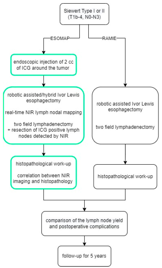
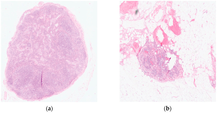
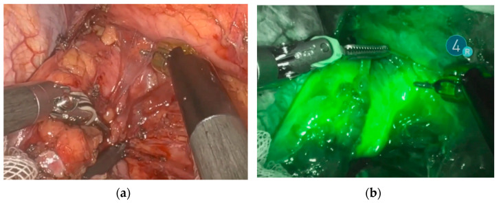
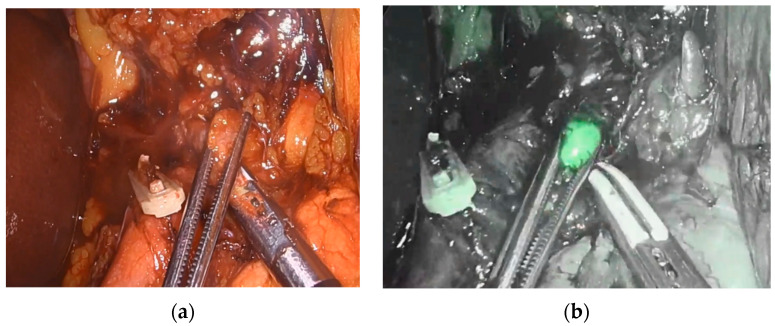
While the sentinel lymph node concept is routinely applied in other surgical fields, no established and valid modality for lymph node mapping for esophageal cancer surgery currently exists. Near-infrared light fluorescence (NIR) using indocyanine green (ICG) has been recently proven to be a safe technology for peritumoral injection and consecutive lymph node mapping in small surgical cohorts, mostly without the usage of robotic technology. The aim of this study was to identify the lymphatic drainage pattern of esophageal cancer during highly standardized RAMIE and to correlate the intraoperative images with the histopathological dissemination of lymphatic metastases. Patients with clinically advanced stage squamous cell carcinoma or adenocarcinoma of the esophagus undergoing a RAMIE at our Center of Excellence for Surgery of the Upper Gastrointestinal Tract were prospectively included in this study. Patients were admitted on the day prior to surgery, and an additional EGD with endoscopic injection of the ICG solution around the tumor was performed. Intraoperative imaging procedures were performed using the Stryker 1688 or the FIREFLY fluorescence imaging system, and resected lymph nodes were sent to pathology. A total of 20 patients were included in the study, and feasibility and safety for the application of NIR using ICG during RAMIE were shown. NIR imaging to detect lymph node metastases can be safely performed during RAMIE. Further analyses in our center will focus on pathological analyses of ICG-positive tissue and quantification using artificial intelligence tools with a correlation of long-term follow-up data.
虽然前哨淋巴结概念在其他外科领域已常规应用,但目前食管癌手术尚无既定且有效的淋巴结定位方法。使用吲哚菁绿(ICG)的近红外光荧光(NIR)最近已被证明是一种安全的技术,可用于在小型手术队列中进行肿瘤周围注射及连续的淋巴结定位,多数情况下无需使用机器人技术。本研究的目的是确定在高度标准化的机器人辅助微创食管癌根治术(RAMIE)中食管癌的淋巴引流模式,并将术中图像与淋巴结转移的组织病理学扩散情况相关联。在我们的上消化道手术卓越中心接受RAMIE的临床晚期食管鳞状细胞癌或腺癌患者被前瞻性纳入本研究。患者在手术前一天入院,并额外进行了一次内镜下肿瘤周围注射ICG溶液的内镜检查(EGD)。术中成像程序使用史赛克1688或FIREFLY荧光成像系统进行,切除的淋巴结送病理检查。本研究共纳入20例患者,结果显示在RAMIE期间使用ICG进行NIR的应用具有可行性和安全性。在RAMIE期间可以安全地进行NIR成像以检测淋巴结转移。我们中心的进一步分析将集中于ICG阳性组织的病理分析以及使用人工智能工具进行定量分析,并与长期随访数据相关联。