Department of Biomedical Sciences, Seoul National University College of Medicine, 103 Daehak-Ro, Jongro-Gu, Seoul, 03080, Korea.
Department of Biochemistry, Molecular Biology and Biophysics, University of Minnesota, Minneapolis, MN, 55455, USA.
Acta Neuropathol Commun. 2023 May 20;11(1):83. doi: 10.1186/s40478-023-01575-0.
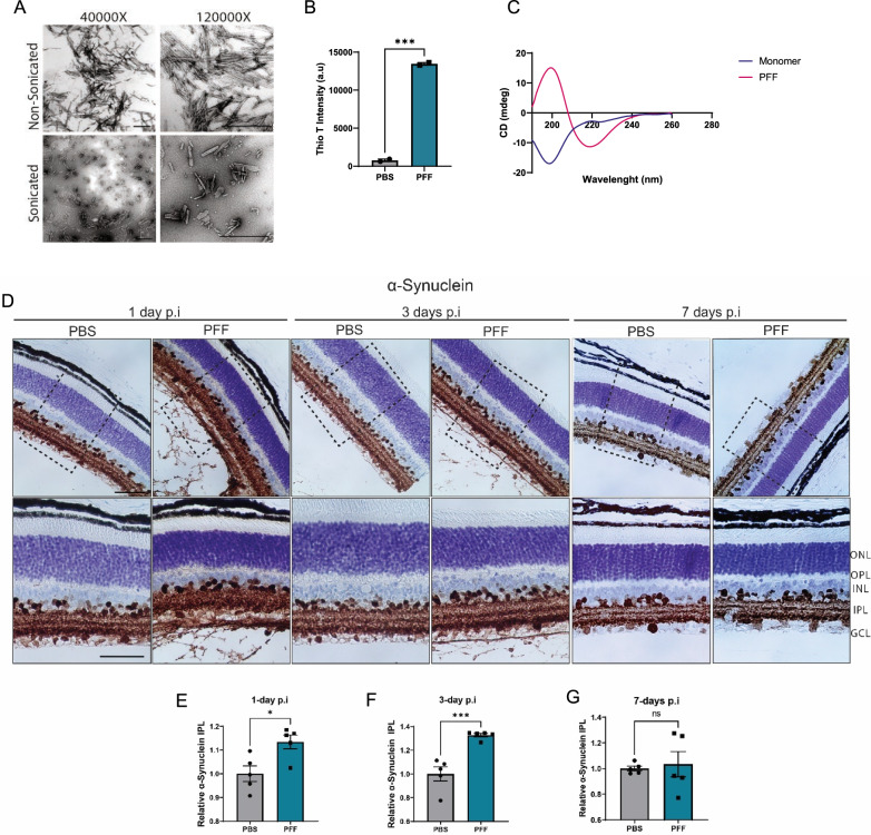
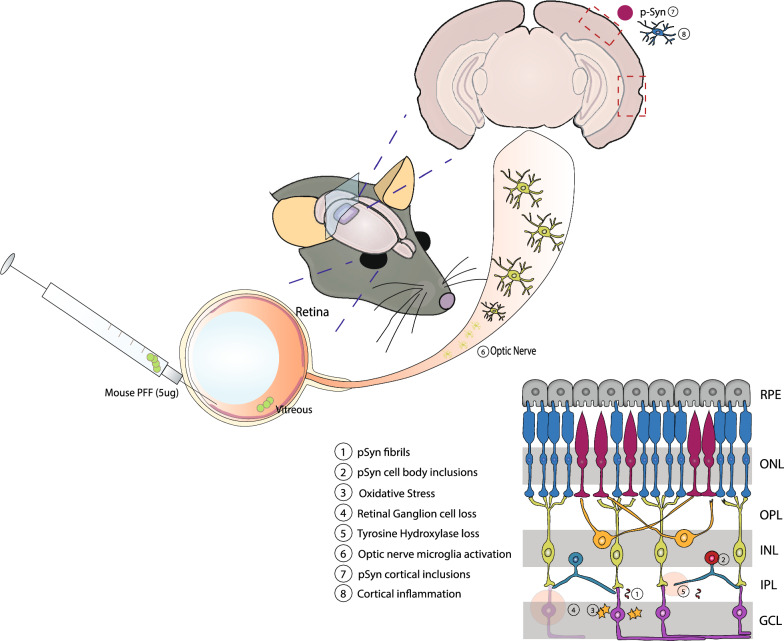
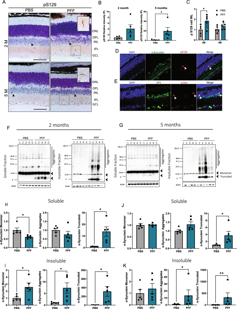
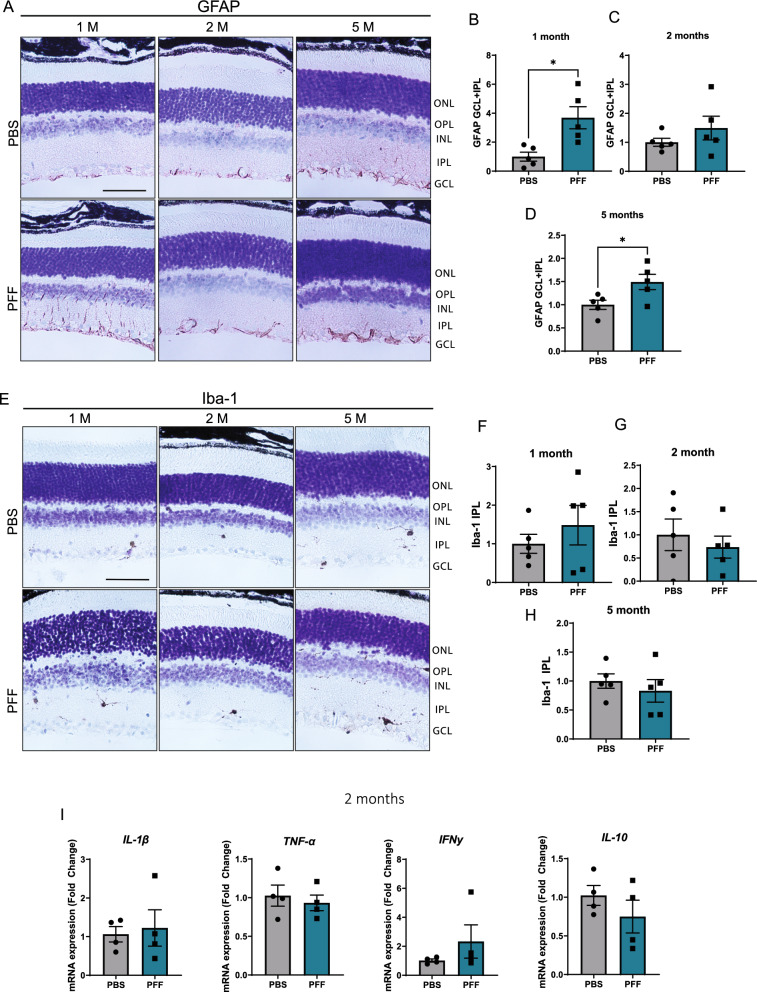
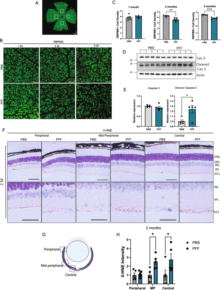
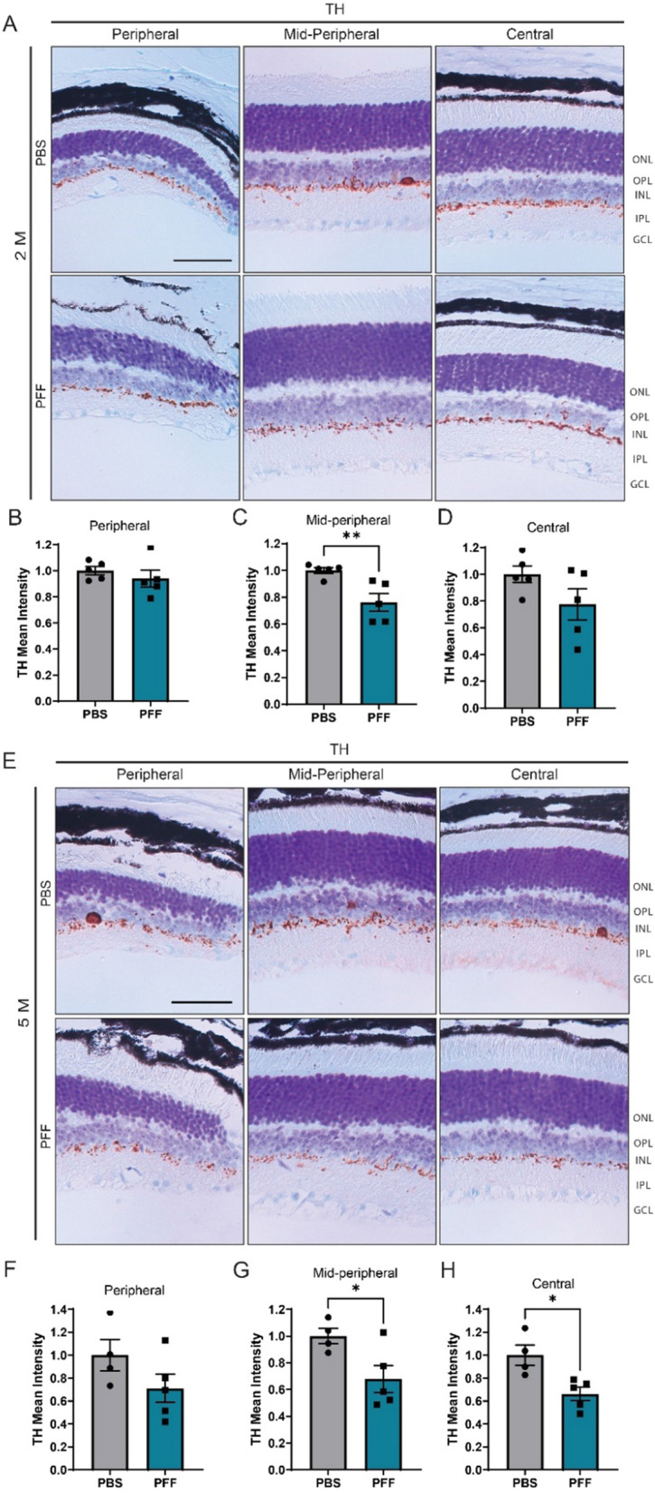
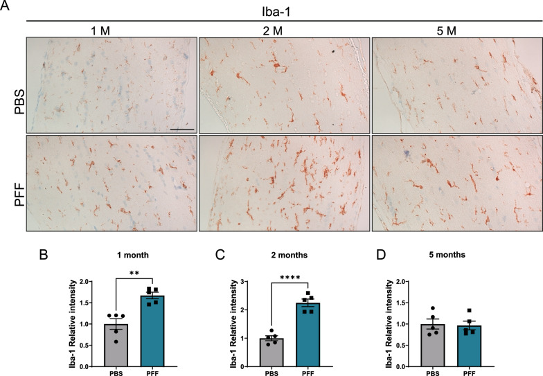
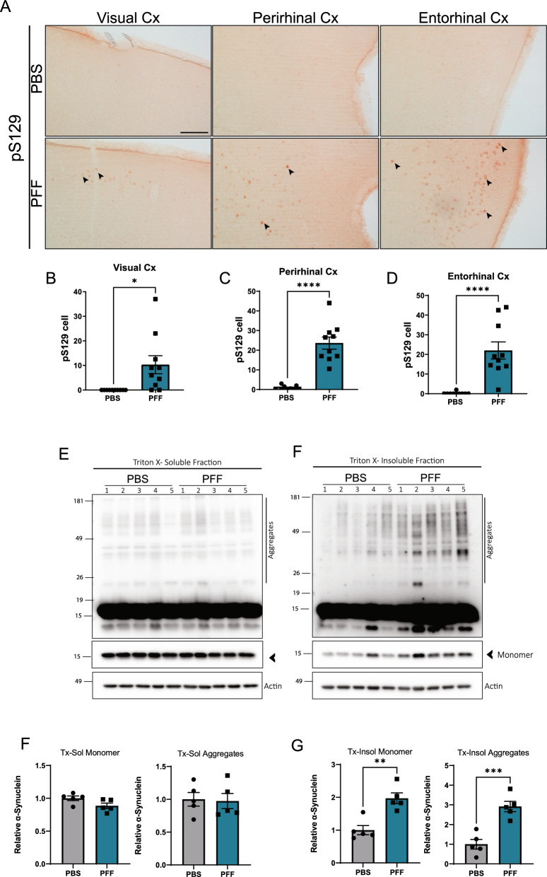
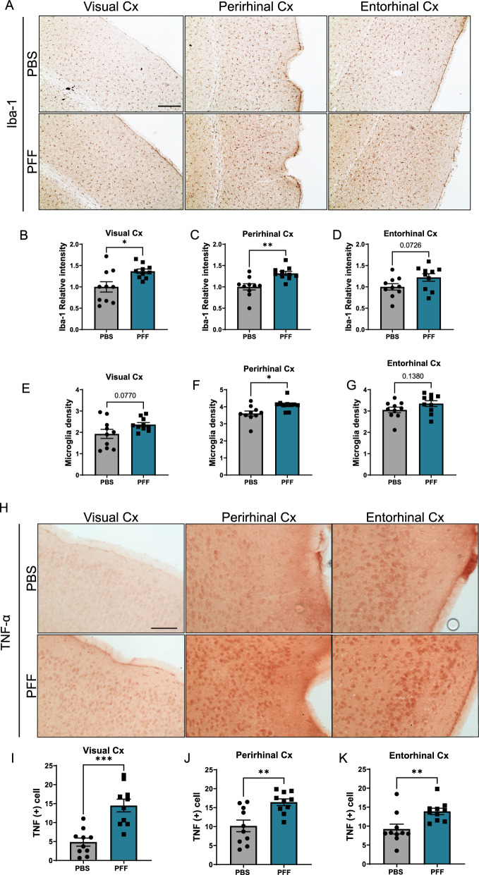
Parkinson's disease (PD) is a neurodegenerative disorder characterized by the aggregation of misfolded α-synuclein and progressive spreading of the aggregates from a few discrete regions to wider brain regions. Although PD has been classically considered a movement disorder, a large body of clinical evidence has revealed the progressive occurrence of non-motor symptoms. Patients present visual symptoms in the initial stages of the disease, and accumulation of phospho-α-synuclein, dopaminergic neuronal loss, and retinal thinning has been observed in the retinas of PD patients. Based on such human data, we hypothesized that α-synuclein aggregation can initiate in the retina and spread to the brain through the visual pathway. Here, we demonstrate accumulation of α-synuclein in the retinas and brains of naive mice after intravitreal injection of α-synuclein preformed fibrils (PFFs). Histological analyses showed deposition of phospho-α-synuclein inclusions within the retina 2 months after injection, with increased oxidative stress leading to loss of retinal ganglion cells and dopaminergic dysfunction. In addition, we found accumulation of phospho-α-synuclein in cortical areas with accompanying neuroinflammation after 5 months. Collectively, our findings suggest that retinal synucleinopathy lesions initiated by intravitreal injection of α-synuclein PFFs spread to various brain regions through the visual pathway in mice.
帕金森病(PD)是一种神经退行性疾病,其特征是错误折叠的α-突触核蛋白聚集以及聚集体从少数离散区域向更广泛的大脑区域进行渐进性扩散。尽管 PD 通常被认为是一种运动障碍,但大量临床证据表明非运动症状会逐渐出现。患者在疾病的初始阶段出现视觉症状,并且在 PD 患者的视网膜中已经观察到磷酸化α-突触核蛋白、多巴胺能神经元丧失和视网膜变薄的现象。基于这些人类数据,我们假设α-突触核蛋白的聚集可以从视网膜开始,并通过视觉通路传播到大脑。在这里,我们通过向视网膜内注射α-突触核蛋白原纤维(PFF)证明了α-突触核蛋白在未受感染的小鼠的视网膜和大脑中的积累。组织学分析显示,注射后 2 个月,视网膜内沉积有磷酸化α-突触核蛋白包涵体,随之而来的氧化应激导致视网膜神经节细胞丧失和多巴胺能功能障碍。此外,我们发现 5 个月后皮质区域内积累了磷酸化α-突触核蛋白,同时伴有神经炎症。总之,我们的研究结果表明,通过向视网膜内注射α-突触核蛋白 PFF 引发的视网膜突触核蛋白病病变可以通过视觉通路在小鼠体内扩散到各种大脑区域。