Department of Ophthalmology, Medical Faculty Mannheim, Heidelberg University, Mannheim, Germany.
Institute of Molecular and Clinical Ophthalmology Basel, Basel, Switzerland.
Invest Ophthalmol Vis Sci. 2023 May 1;64(5):25. doi: 10.1167/iovs.64.5.25.
To search for histologic differences in the beta zone between myopic eyes versus eyes with secondary angle-closure glaucoma.
The histomorphometric study consisted of human eyes enucleated due to uveal melanomas or secondary angle-closure glaucoma.
The study included 100 eyes (age: 62.1 ± 15.1 years; axial length: 25.6 ± 3.1 mm; range: 20.0-35.0 mm). In non-highly myopic glaucomatous eyes compared with non-highly myopic nonglaucomatous eyes, the parapapillary alpha zone was longer (223 ± 168 µm vs. 125 ± 128 µm; P = 0.03), beta zone prevalence (15/20 vs. 6/41; P < 0.001) and length (277 ± 245 µm vs. 44 ± 150 µm; P = 0.001) were higher, and RPE cell density in the alpha zone and alpha zone border was lower (all P < 0.05). In highly myopic nonglaucomatous eyes compared with non-highly myopic glaucomatous eyes, parapapillary RPE drusen prevalence (2/19 vs. 10/10; P = 0.01) and alpha zone prevalence (2/19 vs. 16/20; P < 0.001) and length (23 ± 68 µm vs. 223 ± 168 µm; P < 0.001) were lower. In non-highly myopic glaucomatous eyes, Bruch's membrane (BM) thickness decreased (P < 0.001) from the beta zone (6.0 ± 3.1 µm) to the alpha zone (5.1 ± 4.3 µm) and peripheral to it (3.0 ± 0.9 µm). In highly myopic nonglaucomatous eyes, BM thickness did not differ (P > 0.10) between all three regions. In the total study population, RPE cell density in the alpha zone (24.5 ± 9.3 cells/240 µm) was higher than at the alpha zone border (19.2 ± 4.8 cells/240 µm; P < 0.001) or peripheral to it (19.0 ± 3.6 cells/240 µm; P < 0.001).
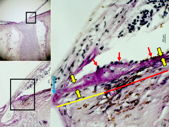
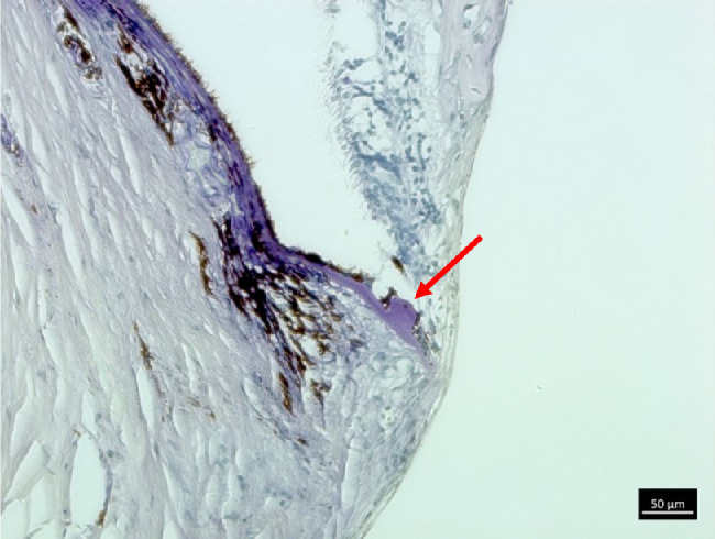
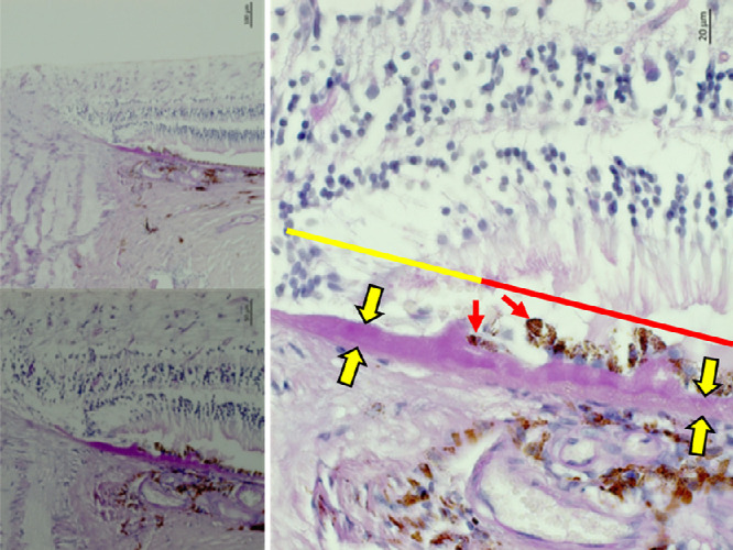
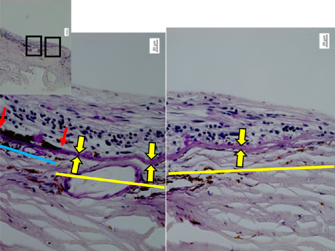
The glaucomatous beta zone in eyes with chronic angle-closure glaucoma (with the alpha zone, parapapillary RPE drusen, thickened BM, and higher RPE cell count in the adjacent alpha zone) differs histologically from the myopic beta zone (characterized by the absence of the alpha zone and parapapillary RPE drusen, unremarkable BM thickness, and unremarkable parapapillary RPE). The differences suggest different etiologies of the glaucomatous versus myopic beta zone.
寻找近视眼中β区与继发性闭角型青光眼眼中β区的组织学差异。
本组织形态计量学研究包括因葡萄膜黑色素瘤或继发性闭角型青光眼而被摘除眼球的人类眼球。
该研究共纳入 100 只眼球(年龄:62.1±15.1 岁;眼轴长度:25.6±3.1mm;范围:20.0-35.0mm)。与非高度近视性青光眼眼相比,非高度近视性非青光眼眼中视盘旁α 区较长(223±168μm 比 125±128μm;P=0.03),β区更常见(15/20 比 6/41;P<0.001)且更长(277±245μm 比 44±150μm;P=0.001),α 区和α 区边界的 RPE 细胞密度更低(均 P<0.05)。与非高度近视性青光眼眼相比,高度近视性非青光眼眼中视盘旁 RPE 玻璃疣的发生率(2/19 比 10/10;P=0.01)和α 区的发生率(2/19 比 16/20;P<0.001)和长度(23±68μm 比 223±168μm;P<0.001)均较低。在非高度近视性青光眼眼中,Bruch 膜(BM)厚度从β区(6.0±3.1μm)到α区(5.1±4.3μm)和其外周(3.0±0.9μm)逐渐变薄(P<0.001)。在高度近视性非青光眼眼中,所有三个区域的 BM 厚度均无差异(P>0.10)。在总研究人群中,α 区的 RPE 细胞密度(24.5±9.3 个细胞/240μm)高于α 区边界(19.2±4.8 个细胞/240μm;P<0.001)或其外周(19.0±3.6 个细胞/240μm;P<0.001)。
慢性闭角型青光眼眼中的青光眼β区(伴有α区、视盘旁 RPE 玻璃疣、增厚的 BM 和相邻α区中更高的 RPE 细胞计数)在组织学上与近视眼中的β区不同(其特征为缺乏α区和视盘旁 RPE 玻璃疣、BM 厚度无明显变化以及视盘旁 RPE 无明显变化)。这些差异表明青光眼β区与近视β区的发病机制不同。